Unit 1 · Contents
Biological Bases of Behavior
From inherited tendencies to the messages that let you think, sleep, sense, and act.
Watch Unit 1 Review
Get Psyched · Unit 1 video review
Open on YouTube If the player is blocked, open the review directly.
One Connected Biological Story
Every thought, sensation, and action depends on biological systems. Unit 1 connects inherited predispositions and experience with the mechanisms that make behavior possible: a communicating nervous system, specialized but connected brain networks, changing sleep states, and sensory systems that convert the world into neural information.
Begin at the broadest level. Heredity and environment influence how people develop. The nervous system then carries and coordinates messages. Neurons explain communication at the cellular level; the brain explains how connected networks regulate, interpret, remember, and act. Sleep changes the conditions under which these networks operate. Sensation supplies the input that later cognitive processes organize and interpret.
The central skill is explaining a functional connection. When you encounter a scenario, identify the relevant detail, name the process, and show why that process accounts for the described behavior. A label alone is insufficient. Ask where a message enters, how it travels, what changes it, and what consequence follows. The original worked examples throughout this guide model that reasoning.
Biology supplies part of an explanation, not a verdict about a person. Brain functions overlap; chemical effects depend on pathways; study designs limit conclusions. Use the comparisons and diagrams to follow mechanisms without assuming that one structure, hormone, or inherited trait explains everything.
After each worked example, explain the connection in your own words. Use the vocabulary reference to return to an explanation, then compare the nearest competing concept.
Explore the Labs
Connect brain function, neural communication, sleep and sensation through interactive experiences.
Neuron Lab
Follow neural signaling, synaptic communication and a reflex pathway.
Topic 1.4Brain Lab
Explore brain structures and connect their locations with their functions.
Topic 1.5Sleep Cycle Explorer
Move through an illustrative night and compare sleep stages.
Topic 1.6Eye Lab
Explore the eye, focus an image and trace visual signaling.
Topic 1.6Ear Lab
Follow sound through the ear and explore hearing and balance.
Topic 1.6Sensory Threshold Workbench
Compare tone detection across trials and differences between weights.
Heredity & Environment
Interaction of Heredity and Environment
Why do people differ? Biology and experience work together. The useful question is how those influences interact, rather than which one wins.
CED printed p. 34Predispositions Meet Experience
Heredity, often called nature, consists of inherited or biologically predisposed characteristics that influence physical traits, behavior, and mental processes. A predisposition makes an outcome more likely under some conditions; it does not make the outcome inevitable. Environment, often called nurture, includes experiences outside the individual, such as family relationships, education, opportunities, nutrition, and cultural expectations.
These influences interact. An inherited tendency toward high activity may affect which activities a child seeks; access to a safe playground may affect how that tendency develops. Experience also changes the biological systems involved in learning. You cannot explain a person by assigning every behavior to a separate nature box or nurture box. The same behavior can reflect both.
A biological explanation describes mechanisms; it does not establish that a trait is unchangeable or that differences between people justify differences in their rights. For this course, learn the relationship between heredity and experience. Detailed genetic mechanisms are outside the exam scope.
The same behavior can reflect both the capacities a learner brings and the experiences available to that learner.
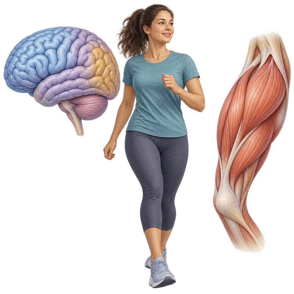
Biological Predispositions
Influence the tendencies and capacities brought to an experience.
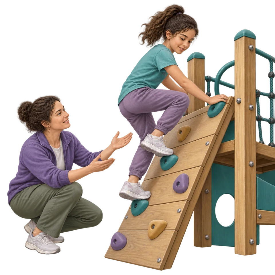
Experiences and Opportunities
Influence how tendencies and capacities develop and are expressed.
Heredity ↔ environment: interacting influences on behavior and mental processes.
An Evolutionary Explanation Is a Testable Proposal
The evolutionary perspective asks how natural selection could have favored tendencies that supported survival and reproductive success across generations. In natural selection, heritable variations associated with greater reproductive success can become more common in a population. Individuals do not acquire an adaptation simply because they need it, and an evolutionary account does not mean every current behavior is useful in every current environment.
For example, a tendency to attend rapidly to possible threats could be interpreted as having helped ancestors avoid danger. That is an original teaching illustration of the kind of explanation this perspective proposes, not a published finding about a specific person. An explanation still needs evidence; a plausible story alone is insufficient.
Eugenics refers to attempts to control reproduction according to judgments about supposedly desirable inherited traits. People have misused evolutionary and hereditary ideas to support discrimination and coercion. Describing natural selection does not justify ranking human worth or treating social disadvantage as evidence of inherited inferiority.
What Twin, Family, and Adoption Studies Can Tell Us
Behavioral genetic researchers compare patterns of similarity among people whose biological relationships and environments differ. Twin studies often compare identical and fraternal twins, or examine twins raised in different environments. Greater similarity among more closely genetically related twins can support a hereditary contribution, but twins also differ in how similarly they are treated and in the environments they experience.
Family studies examine whether traits cluster among relatives and how similarity changes with degree of biological relationship. Families share experiences as well as heredity, so a family pattern cannot by itself separate those influences. Adoption studies compare adopted people with biological relatives and with adoptive relatives. Resemblance to biological relatives can suggest hereditary contributions; resemblance to adoptive relatives can suggest environmental contributions. Prenatal experiences, selective placements, and contact among relatives can complicate either inference.
These comparisons are generally non-experimental: researchers do not randomly assign inherited characteristics or family histories. A pattern of resemblance is evidence about an association, not proof that a particular gene causes a behavior. Use converging evidence and explain limitations instead of treating one study design as a nature-versus-nurture verdict.
| Design | Comparison | Key limit |
|---|---|---|
Twin | Different degrees of genetic similarity and/or rearing environments | Key limitGenetic similarity and similarity of treatment may vary together. |
Family | Traits across biological relatives | Key limitRelatives often share environments. |
Adoption | Biological and adoptive relatives | Key limitPlacement and prenatal experience are not randomly assigned. |
Nervous System
Overview of the Nervous System
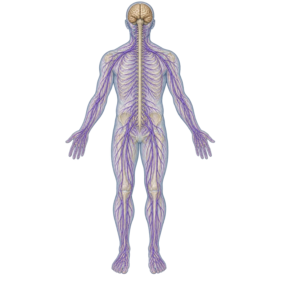
The nervous system connects sensory input, coordination, and action. Its subdivisions describe different jobs within one communicating system.
CED printed p. 35One Network, Several Kinds of Control
The central nervous system (CNS) consists of the brain and spinal cord. It receives and integrates information and helps coordinate responses throughout the body. The spinal cord also coordinates some rapid responses, so central does not mean that every response must wait for conscious thought in the brain.
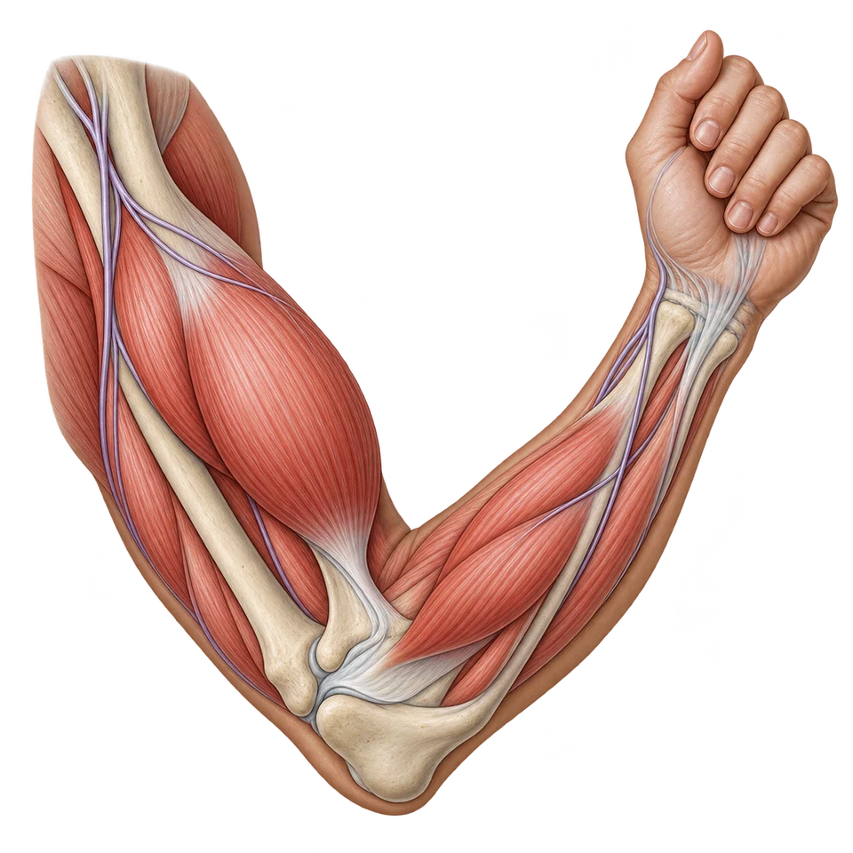
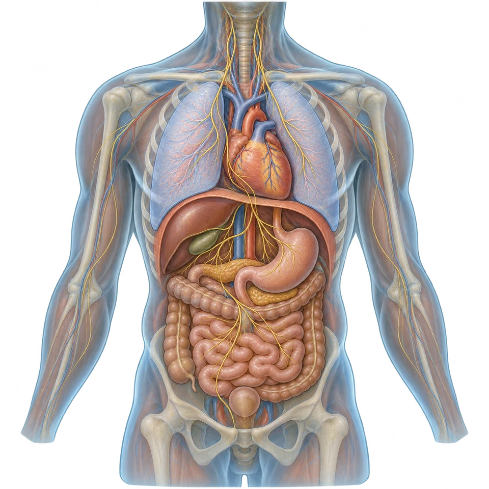
The peripheral nervous system (PNS) links the CNS with the rest of the body. Incoming signals provide information about the body and environment; outgoing signals influence muscles and organs. The PNS includes the somatic nervous system, associated with voluntary skeletal-muscle movement, and the autonomic nervous system, which regulates largely involuntary functions such as heart rate and digestion. Somatic pathways also participate in involuntary reflexes; voluntary is a useful functional distinction, not a rule that every somatic response is consciously chosen.
The autonomic system includes the sympathetic division, which mobilizes resources for action, and the parasympathetic division, which supports maintenance, digestion, and recovery. During an urgent challenge, sympathetic activity can increase heart rate and redirect resources. After the challenge, parasympathetic activity helps restore a calmer pattern. Both divisions continually regulate the body; they are not two switches that completely turn one another off.
Read the whole system first, then follow its membership branches. Central and peripheral are the two main divisions. Only the peripheral branch contains the somatic and autonomic divisions; only the autonomic branch contains sympathetic and parasympathetic.
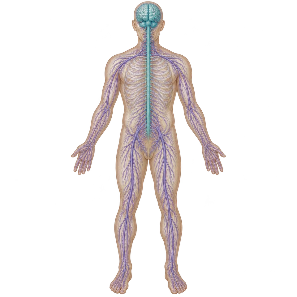
Nervous System
One communication system, two major divisions.
Two main divisions
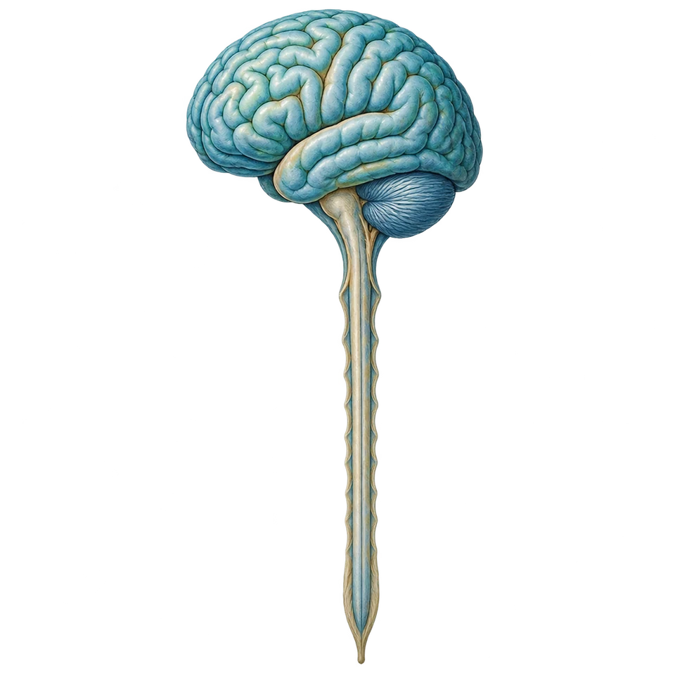 Part of Nervous system
Central Nervous System · CNS
Brain + spinal cord. Integration and coordination.
 Part of Nervous system
Peripheral Nervous System · PNS
Links CNS and body. Carries incoming and outgoing messages.
Within PNS · two divisions
 Part of PNS
Somatic
Skeletal-muscle control. Reflex pathways need not involve a deliberate choice.
 Part of PNS
Autonomic
Largely involuntary regulation of internal functions.
Within autonomic · two branches
 Part of Autonomic
Sympathetic
Mobilizes resources. The activity scene illustrates bodily preparation for demand.
 Part of Autonomic
Parasympathetic
Supports maintenance and recovery. The meal scene illustrates digestive maintenance.
Neurons & Neural Firing
The Neuron and Neural Firing
Behavior depends on communication within and between cells. Learn the sequence and the consequences of changing it, rather than treating the neuron as a labeling exercise.
CED printed pp. 36–38Cells Support Communication; Reflexes Coordinate a Response
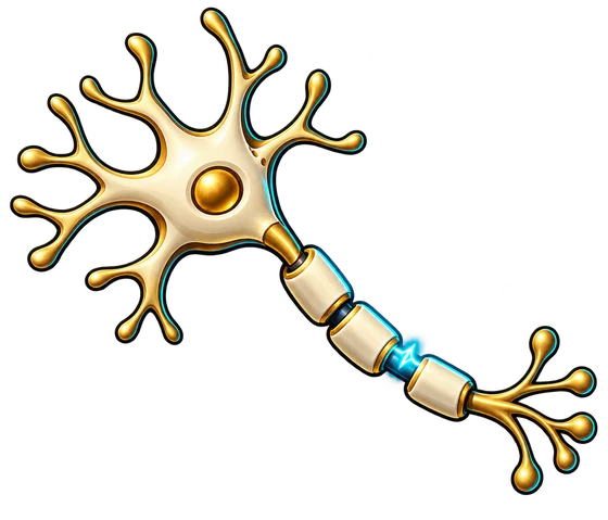
Neurons are specialized cells that transmit information. Dendrites receive many incoming signals; the cell body supports the cell and integrates input; an axon carries an action potential toward terminals that communicate with another cell. These labels help explain the process. They are not a separate demand to memorize an unlabeled anatomical picture.
Glial cells support the nervous system by providing structure, insulation, communication support, and waste transport. Some glia form myelin, insulating material around axons that helps signals travel efficiently. Glia are active partners in neural functioning, not merely filler between neurons.
In a reflex arc, a sensory neuron carries information from a receptor toward the spinal cord. An interneuron within the CNS links and processes signals in a typical withdrawal reflex. A motor neuron carries an outgoing signal to a muscle. Touching a very hot surface can therefore trigger withdrawal before conscious awareness catches up; information also travels to the brain. Sensory describes incoming information, motor describes outgoing action, and interneuron describes connections within the CNS.
Neuron Lab
Explore the Structure. Follow the Signal.
Explore the structures, compare inputs, follow chemical communication, and trace one withdrawal pathway.
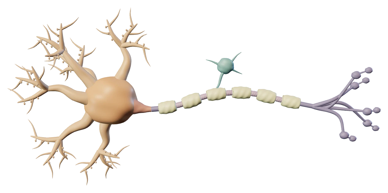
Choose a structure or select it directly on the model. Use the view buttons to rotate and zoom.
Neuron Lab: Complete 2D Reference
A Representative Myelinated Multipolar Neuron. Explore the structures, compare inputs, follow chemical communication, and trace one withdrawal pathway.
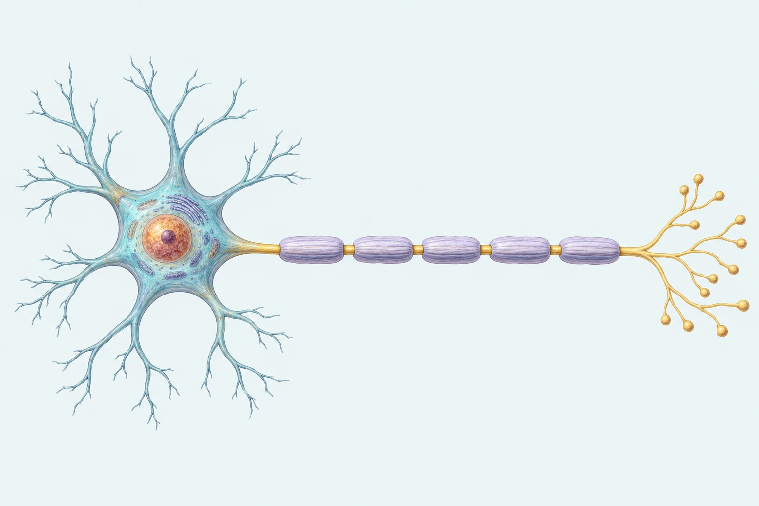
Follow input from dendrites into the cell body, then follow the single axon toward its terminal branches. Lavender sleeves depict myelin around the axon; the visible golden gaps belong to that same continuous axon.
- 1. Dendrites
- Branching input extensions at the left receive incoming signals.
- 2. Cell body
- The soma supports the neuron and integrates input; the callout points to its cytoplasm.
- 3. Axon
- The continuous golden axon carries an action potential toward the terminals. This callout points to an exposed gap between sleeves.
- 4. Myelin
- Segmented insulating sleeves surround the same axon and support efficient signaling.
- 5. Axon terminals
- The branching end at right communicates with other cells through terminal boutons.
Solid arrow: action potential travels along the axon toward the terminals.
Explore the Neuron
- Dendrites
- Function: Receive many incoming signals from other cells.
Location: The branching processes around the cell body. Many branches provide places for input; the cell combines those effects. This is one representative multipolar neuron; neuronal forms vary.
- Cell Body (Soma)
- Function: Supports the cell and helps integrate its inputs.
Location: The central body joining the branches and the start of the axon. Its role connects the incoming branches to the outgoing pathway. The nucleus supports the cell; it is not a switch that launches every impulse.
- Axon Beginning
- Function: A common site where sufficient combined input initiates an action potential.
Location: The initial segment just beyond the soma and before the first myelin segment. This is the marked recording location for the signal graph. The model identifies this neuron’s initiation region; other forms can differ.
- Axon
- Function: Carries an action potential toward the terminal branches.
Location: The continuous fiber passing through the myelin segments and exposed gaps. The electrical change is renewed along the fiber; it is not neurotransmitter flowing down a tube. The fiber is shortened and enlarged for viewing.
- Myelin
- Function: Glial insulation helps signals travel efficiently.
Location: The separate wraps around portions of the continuous axon. Electrical change spreads beneath a wrap and triggers a renewed action potential at the next gap. Glia also provide structure, communication support and waste transport; this model highlights insulation.
- Gaps Between Myelin
- Function: Renew the action potential as it moves along the myelinated axon.
Location: Short regions of axon between adjacent myelin segments. The gap is an exposed part of the same axon, not a break that an impulse jumps across. Gap sizes and spacing are expanded for clarity.
- Terminal Branches
- Function: Communicate with other cells through synaptic contacts.
Location: The small branches and boutons at the end of the axon. Electrical arrival can trigger the release of chemical messengers at the contact explored next. The close-up shows one representative chemical synapse.
- Glial Support
- Function: Supports and insulates nearby neural tissue.
Location: The small support-cell cue and its connection to a visible wrap. Myelin comes from glial cells; it is not a decorative sleeve. One visible process does not represent every glial type, function or myelin segment.
Send a Signal
Relative teaching scale: 0–100; this example reaches threshold at 60. Below threshold, the local input dissipates and no action potential propagates. At or above threshold, this single-event example has the same full spike height.
- Resting State (0 ms; -70 mV at the recording site). At the marked axon beginning, the inside is relatively negative. The input has not yet arrived. Resting potential is a difference across this membrane, not a signal moving down the axon. Refractory context: ready.
- Threshold Reached (0.5 ms; -55 mV at the recording site). Combined input brings this example’s recording site to threshold. A full action potential begins. The input is sufficient to initiate the event; reaching threshold does not set a continuously adjustable spike height. Refractory context: beginning.
- Depolarization (0.8 ms; 30 mV at the recording site). At the recording site, the membrane becomes less negative and briefly positive. This is the peak of the local action potential. The same spike occurs for every suprathreshold input in this single-event example. Refractory context: cannot initiate another impulse.
- Repolarization (1.3 ms; -40 mV at the recording site). The recording site returns toward its resting voltage after the peak. This falling voltage is a recovery phase, not a smaller action potential or a new threshold. Refractory context: recovering.
- Brief Undershoot (2.1 ms; -80 mV at the recording site). The local membrane is briefly more negative than it was at rest. During recovery it can temporarily be harder to initiate another impulse. Refractory context: temporarily harder to fire.
- Return to Rest (3 ms; -70 mV at the recording site). The recording site returns to this example’s resting potential. Electrical recovery at this site is separate from transmitter cleanup at a synapse. Refractory context: ready again.
- Along the Myelinated Axon (4 ms; -70 mV at the recording site). Electrical change spreads beneath each insulating segment and renewed action potentials appear at the gaps. The axon is continuous. Myelin improves propagation without carrying chemical messengers or creating empty breaks. Refractory context: recording site ready.
- Arrival at the Terminal (5 ms; -70 mV at the recording site). The impulse reaches the terminal branches. The next mode follows chemical communication at one contact. The graph still records the axon beginning, so it remains at rest while the later event occurs elsewhere. Refractory context: recording site ready.
The graph records membrane voltage at the marked axon beginning. The 0–5ms window and intermediate sample values are schematic; they are not measurements of this model or a speed estimate. Replaying or scrubbing revisits a teaching stage rather than reversing physiology.
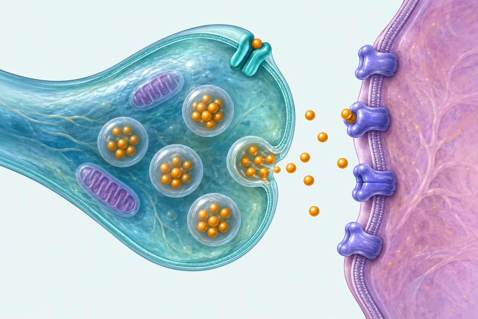
An arriving electrical impulse triggers chemical release at the sending terminal. Transmitter crosses the extracellular gap and binds to receiving-cell receptors. Reuptake is a return pathway into the sending terminal, not the receiving membrane and not the axon’s refractory reset.
- 1. Sending terminal
- The turquoise cutaway is the end of the sending axon. One vesicle is shown merging with its membrane to release transmitter.
- 2. Synaptic gap
- The space between the two cell membranes contains released chemical messengers.
- 3. Receiving membrane
- The lavender cell surface carries embedded receptor proteins; reception changes the receiving cell’s response.
- 4. Receptors
- Purple membrane proteins receive transmitter. The bound amber symbol at the upper-middle receptor illustrates binding.
- 5. Reuptake transporter
- The teal protein in the sending membrane illustrates return of released transmitter into the sender.
- 6. Neurotransmitter
- Amber symbols represent chemical messengers, not literal measured molecular size or a concentration.
Solid arrow: release and movement toward receptor binding.
Dashed arrow: reuptake into the sending membrane’s transporter.
Cross the Synapse
- Sending Terminal
- The arriving electrical impulse can trigger release from vesicles in this terminal. The cutaway bouton connected to the sending axon.
- Synaptic Vesicles
- Store transmitter and fuse with the sending membrane to release it. Inside the sending terminal.
- Synaptic Gap
- The extracellular space through which released transmitter diffuses. Between two distinct cell membranes.
- Receptors
- Recognize transmitter and change the receiving cell’s response. Embedded in the receiving membrane.
- Receiving Membrane
- Its receptors turn chemical interaction into a change in the receiving cell. The surface of another cell, shown as a continuing dendritic patch.
- Reuptake Transporter
- Returns released transmitter into the sender in this selected clearance example. Embedded in the sending membrane, separate from receiver receptors.
- Sending Cell Ready. Two distinct cells meet at this contact. The sending impulse begins above the terminal; the receiving cell rests. Choose a receptor context, then send the same presynaptic action potential in either example.
- Signal Arrives. The electrical impulse reaches the sending terminal. No transmitter has crossed the gap yet. An inhibitory synapse still receives a normal presynaptic action potential.
- Vesicle Fusion and Release. The selected vesicle fuses with the sending membrane and releases individual transmitter molecules. The vesicle membrane remains with the sender; whole vesicles do not cross the gap.
- Across the Gap. Released molecules diffuse through the extracellular gap toward the receiving membrane. This chemical movement between cells follows electrical transmission within the sender.
- Receptor Binding. Some released molecules bind to suitable receptors embedded in the receiving membrane. Receptor interaction changes the receiving cell. It does not guarantee a new action potential.
- Receiving Cell Response. The selected receptor context produces a graded postsynaptic change. The receiving-cell display shows its direction and size. Inputs still combine at the receiving axon beginning. Both examples remain below the firing-threshold reference.
- Clearance and Recovery. Transmitter returns through sender transporters or diffuses away. The receiving response returns to rest in this example. This selected time course is illustrative; different receptors and transmitters have different clearance and response patterns.
In this selected excitatory receptor context, reception makes reaching threshold more likely. Other inputs still matter. In this selected inhibitory receptor context, reception makes reaching threshold less likely. Inhibitory does not mean bad. The comparison changes receptor context; it does not claim the same transmitter has the same effect at every synapse or that the receiver automatically fires.
Receiving Cell Response
The receiving cell becomes less negative and moves toward the threshold reference. This selected response stays below threshold, so no receiving action potential begins. The illustrative peak is −61 mV from −70 mV at rest.
In this hyperpolarizing example, the receiving cell becomes more negative and moves away from the threshold reference. No receiving action potential begins. The illustrative trough is −77 mV from −70 mV at rest. This shows one hyperpolarizing inhibitory response. Inhibition can also reduce the effect of excitatory input without the same voltage dip, called shunting inhibition.
Inputs combine at the receiving axon beginning. This contact’s response does not guarantee a new action potential. Voltage and timing are illustrative; neither example reaches firing threshold. As this example’s message ends, its graded response returns toward rest. Different receptor mechanisms can have different time courses.
Different transmitters use different combinations of uptake, breakdown and diffusion. Reuptake carries transmitter through a membrane transporter into a cell; enzymatic breakdown chemically alters it; diffusion moves it away from the contact. This sequence highlights sender reuptake and diffusion without applying that same cleanup pattern to every transmitter.
Follow a Reflex
Purple Dashed Branch: Information to the Brain · Gold Muscle: Upper-Arm Flexor
- Fingertip Receptor · Receptor. Peripheral nervous system. The illustrated fingertip cue activates incoming sensory information. An example to follow, not a stimulus to try.
- Sensory Pathway · Sensory Neuron. PNS pathway enters CNS. Incoming information travels along the arm toward the cervical spinal cord. Sensory describes incoming information, rather than outgoing muscle activation.
- Cervical Spinal Processing · Interneuron. Central nervous system. A simplified spinal connection helps activate the motor pathway. Information also travels toward the brain. Spinal processing can begin withdrawal before conscious awareness. Information also ascends toward the brain.
- Motor Pathway · Motor Neuron. CNS cell body; PNS axon to muscle. Outgoing information reaches an upper-arm flexor muscle in this example. Motor describes output. The central and peripheral nervous systems work together.
- Elbow Bend and Withdrawal · Responding Muscle. Peripheral target. The upper-arm flexor contracts; the elbow bends and the hand withdraws. Real withdrawal can coordinate several muscles and varies with posture and context.
The shown spinal response does not wait for a conscious decision. The ascending information branch does not gate the motor pathway. A simplified protective pathway, not an exact tract, root or latency map. Other circuits and movements differ.
- 1 · Sensory input
A skin receptor detects a harmful stimulus; a sensory neuron carries the message toward the spinal cord.
- 2 · Central connection
An interneuron in the spinal cord helps connect the incoming signal to the response.
- 3 · Motor output
A motor neuron carries the signal to a muscle that withdraws the hand. The brain also receives information.
From Resting Potential to a Message Across a Synapse
At resting potential, a neuron has an electrical difference across its membrane: the inside is relatively negative. Incoming signals can shift that difference. Excitatory input makes an action potential more likely, and inhibitory input makes it less likely. Inputs combine; a cell does not necessarily fire in response to one incoming message.
When sufficient input reaches the threshold, the neuron generates an action potential, a brief electrical change that travels along the axon. During depolarization, the membrane becomes less negative and briefly reverses its electrical polarity. The all-or-none principle means an individual action potential occurs fully once its threshold is reached; it is not a half-sized impulse for a weak stimulus. Stronger stimulation can change the frequency of firing and the number of neurons recruited, rather than making each impulse bigger.
After firing, the refractory period briefly limits the ability to fire again while the membrane returns toward its resting condition. At an axon terminal, the arriving action potential leads to neurotransmitter release. These chemical messengers cross the synapse, the junction between cells, and bind to receptors on a receiving cell. That receptor activity changes the receiving cell’s likelihood of responding. Electrical signaling along an axon and chemical signaling between cells are different steps.
The message must also end. In reuptake, a released neurotransmitter is taken back into the sending cell. Breakdown or diffusion can also clear some transmitters. Reuptake is a cleanup step at the synapse, not the electrical reset of an axon during its refractory period. The sodium-potassium pump is outside the AP exam scope.
Communication can be disrupted at different places. In multiple sclerosis, damage to myelin in the CNS disrupts efficient signal transmission, potentially affecting movement, sensation, and other functions. In myasthenia gravis, impaired signaling at the nerve-muscle junction, commonly involving acetylcholine receptors, produces muscle weakness. The distinction is damaged axon insulation versus impaired communication to muscle; these are not interchangeable examples of a neuron simply failing to reach threshold.
- 1 · Rest and input
Excitatory and inhibitory signals alter the cell’s electrical state.
- 2 · Threshold and impulse
Reaching threshold initiates a full action potential; depolarization travels along the axon.
- 3 · Release and reception
Terminals release chemical messengers that bind to receptors on the next cell.
- 4 · Clear and reset
Reuptake helps end the synaptic message. The membrane recovers during the refractory period.
Eight Neurotransmitters: Functions Depend on the Pathway
Neurotransmitters carry messages between cells at synapses. Their effects depend on receptors and on where they act. A transmitter may influence several functions, and a complex behavior depends on many transmitters and networks. Excitatory and inhibitory describe an effect on a receiving cell’s likelihood of firing; they do not mean good and bad, or happy and sad.
Use the eight course-listed transmitters below to explain a functional connection. These are major associations, not a one-chemical explanation of a personality, emotion, or disorder. For example, describing dopamine’s role in movement does not erase its role in reward-related learning.
- Dopamine
- Helps regulate movement, motivation, and reward-related learning. Effects vary across pathways; reward learning involves more than pleasure alone.
- Serotonin
- Contributes to mood regulation, sleep, and other functions. Mood cannot be reduced to the amount of one transmitter.
- Norepinephrine
- Supports alertness, attention, and arousal, including responses to challenges.
- Glutamate
- A major excitatory transmitter involved in learning and memory; excitation makes a receiving cell more likely to fire.
- GABA
- A major inhibitory transmitter; it reduces the likelihood of firing in receiving cells and helps regulate neural activity.
- Endorphins
- Naturally occurring chemical messengers involved in pain reduction and related responses.
- Substance P
- Contributes to transmission of pain-related signals. Contrast it with endorphins, which can reduce pain signaling.
- Acetylcholine
- Supports skeletal-muscle activation and contributes to attention, learning, and memory. Its effect depends on its receptor and location.
Hormonal Messages Travel Through the Body
Hormones are chemical messengers released into the bloodstream that affect cells with suitable receptors. Neural messages can be fast and targeted; hormonal signals often spread more broadly and can last longer. Both influence behavior and mental processes, and the nervous and endocrine systems interact. The distinction is the route of communication, not that one system affects the mind while the other affects only the body.
The course specifies these five hormones. Adrenaline helps mobilize the body, while leptin and ghrelin help regulate hunger-related signals. Melatonin helps signal biological night; it is not an on-off sleep switch. Oxytocin contributes to reproductive functions and some social responses; calling it a universal trust chemical would overstate its effects. Detailed endocrine-gland anatomy is excluded, except for the pituitary connection taught in Topic 1.4.
- Adrenaline
- Also called epinephrine; helps prepare the body for action, including increasing cardiovascular activity.
- Leptin
- Signals energy availability and tends to reduce hunger; associate it with satiety rather than meal initiation.
- Ghrelin
- Helps increase hunger and motivates eating; contrast its hunger signal with leptin’s satiety-related signal.
- Melatonin
- Helps regulate the timing of the sleep/wake cycle by signaling biological night.
- Oxytocin
- Participates in childbirth and milk release and contributes to social bonding in context-dependent ways.
| System | Message route | Application |
|---|---|---|
| Neural | Along neurons and across synapses | Acetylcholine helps a motor neuron activate a muscle. |
| Hormonal | Through blood to receptor-bearing cells | Adrenaline helps prepare several body systems for an urgent response. |
Compare the hormone’s target and function with rapid neural signaling. Hormonal and neural influences can work together.
Changing the Message at a Receptor or During Cleanup
Psychoactive drugs change behavior and mental processes by affecting neural communication. An agonist activates a receptor or enhances the action of a neurotransmitter. An antagonist blocks or reduces a neurotransmitter’s action, often by occupying a receptor without activating it in the same way. A reuptake inhibitor blocks a transporter involved in reabsorbing a transmitter, leaving more of that transmitter available in the synapse for longer.
The CED describes these broadly as encouraging or discouraging firing. For accurate application, follow the affected pathway: an agonist acting on an inhibitory receptor can strengthen inhibition. Agonist means increased receptor action, not universally increased overall brain activity. Likewise, a reuptake inhibitor is not a machine that creates new transmitter or permanently turns a neuron on.
Compare the affected step in neural communication. These are alternative mechanisms, not chronological stages.
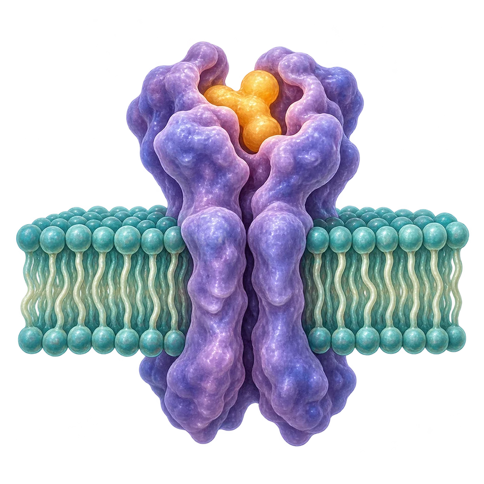
Agonist
Activates a receptor or enhances neurotransmitter action; the effect depends on the receptor and pathway.
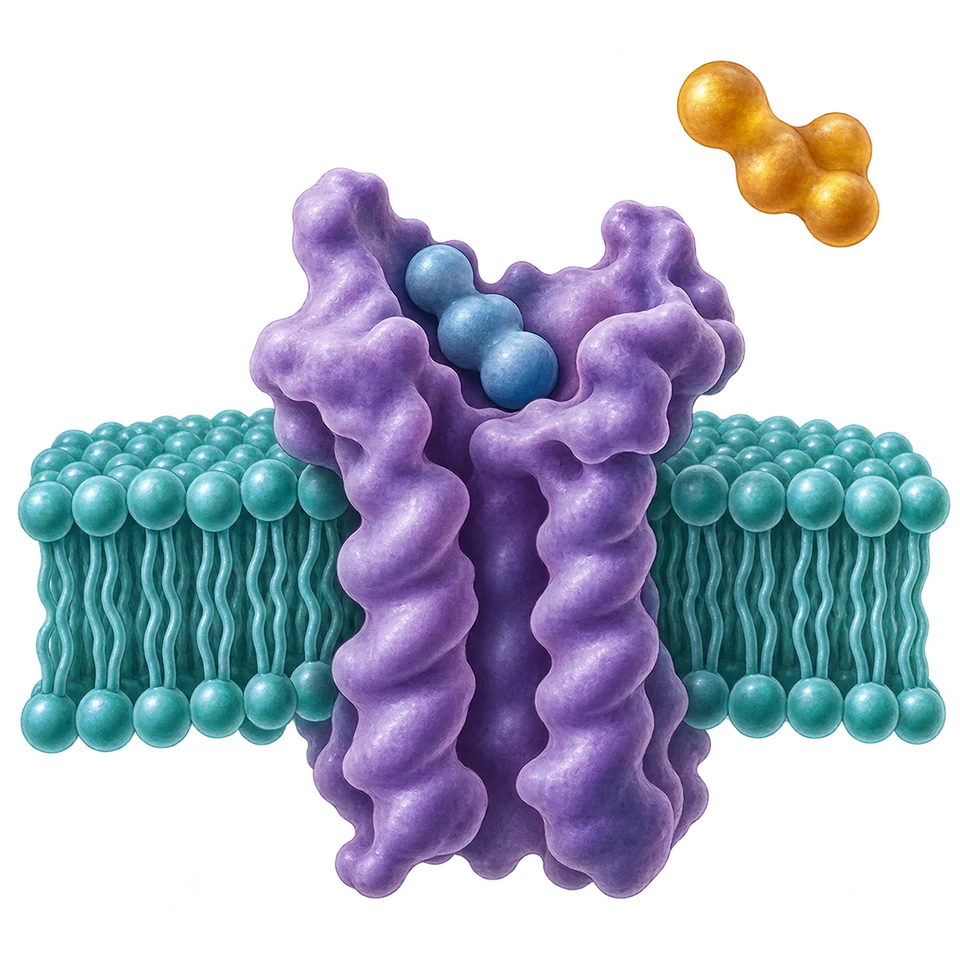
Antagonist
Blocks or reduces neurotransmitter action, often by occupying a receptor without activating it in the same way.
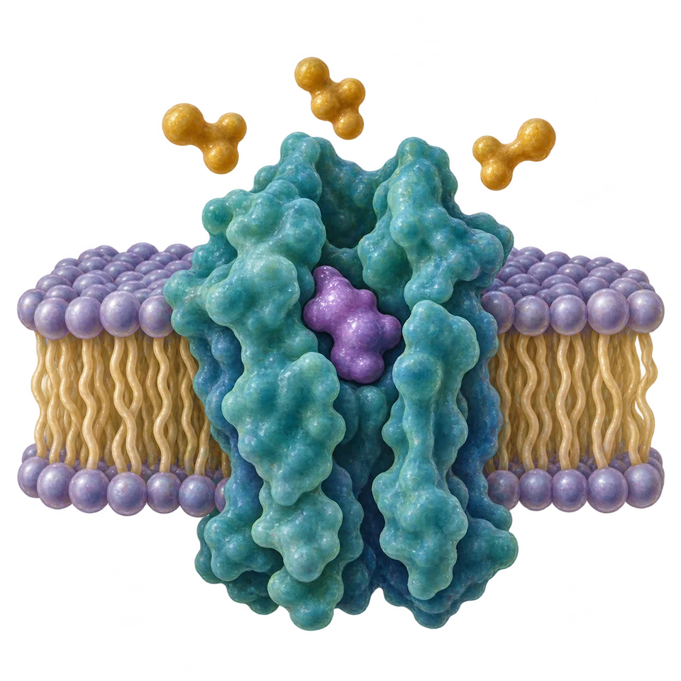
Reuptake Inhibitor
Blocks a transporter involved in transmitter return, leaving more transmitter available in the synapse for longer.
Four Drug Categories and Adaptation to Repeated Use
Drug categories describe typical effects; they are not a guarantee about every dose, person, or setting. Stimulants, such as caffeine and cocaine, generally increase neural activity and arousal. Depressants, such as alcohol, generally reduce neural activity and can impair judgment, coordination, and response speed. Depressant describes activity, not necessarily a sad mood. Alcohol can initially appear to increase sociability because it reduces inhibition.
Hallucinogens alter perception and cognition. The CED uses marijuana as its example in this category; its effects can be varied, so use the course example without assuming that every effect of cannabis is a hallucination. Opioids, such as heroin, act on opioid receptors and commonly reduce pain; they can also strongly affect reward and breathing. A psychological effect concerns experiences such as alertness or perception; a physiological effect concerns bodily functions such as heart rate or motor coordination. A drug can have both.
With tolerance, repeated exposure can reduce the effect of the same dose, so a larger dose may be needed to obtain a previous effect. Addiction involves compulsive use despite harmful consequences. Withdrawal refers to adverse symptoms when a substance on which the body has become dependent is reduced or stopped. Tolerance, dependence, and addiction are related but distinct: needing more for the same effect does not by itself establish compulsive use, and withdrawal describes what happens during reduction or cessation.
| Category | CED example | Typical functional effect |
|---|---|---|
Stimulant | Caffeine; cocaine | Increased activity and arousal |
Depressant | Alcohol | Reduced activity; impaired coordination and judgment |
Hallucinogen | Marijuana | Altered perception and cognition |
Opioid | Heroin | Pain relief; effects on reward and bodily regulation |
The Brain
The Brain
The brain contains specialized regions that work in connected networks. Explain how a function relates to behavior, while avoiding the idea that a complex behavior lives in one isolated spot.
CED printed pp. 39–40Basic Regulation, Alertness, Reward, and Coordination
The brain stem connects the brain with the spinal cord and supports basic functions. Its medulla helps regulate breathing and heart rate, explaining why damage there can endanger basic bodily functioning even when a person’s knowledge is not the primary issue.
The reticular activating system helps regulate alertness and the level of wakefulness. Related brainstem circuits contribute to eye movement and some movement functions. The brain’s reward system is a network that participates in motivation, learning, cognition, and emotion by helping organisms respond to rewarding outcomes. Calling it a reward center is course shorthand; it is not one tiny pleasure button responsible for every rewarding experience.
The cerebellum, at the lower rear of the brain, helps coordinate movement and balance and contributes to procedural learning. A person whose cerebellar function is impaired might have difficulty making smooth, accurately timed movements, even if the decision to move and the ability to produce muscle force remain. Compare regulation of basic bodily activity, maintaining alertness, and coordinating movement rather than treating these structures as interchangeable.
The Cortical Lobes Organize Connected Kinds of Processing
The cerebral cortex is the folded outer layer of the cerebrum, which is divided into left and right hemispheres. The hemispheres communicate through the corpus callosum, a large bundle of connecting fibers. Each hemisphere has frontal, parietal, temporal, and occipital lobes. The cortex also works with connected deeper structures discussed below; those structures should not all be drawn as parts of the outer cortical sheet.
At the front, the frontal lobes participate in planning, linguistic processing, and voluntary movement. The prefrontal cortex supports executive functions such as planning, inhibiting an impulsive response, and organizing actions toward a goal. The motor cortex, near the rear of the frontal lobe, helps direct skeletal-muscle movements, largely on the opposite side of the body.
The parietal lobes, near the upper rear portion of the cortex, contribute to integrating and organizing sensory information. The somatosensory cortex receives and processes bodily touch information. Association areas combine information and support higher functions; they are distributed across the cortex, so do not interpret the parietal association role as the only place where information is integrated.
The temporal lobes, on the sides of the brain, contribute to hearing and language processing. The occipital lobes, at the back, are strongly involved in visual processing. These associations help predict possible consequences of injury, but real effects depend on the specific location, extent of damage, and connected networks.
Brain Lab
A Closer Look at Structure and Function.
Anatomical teaching model · not an activity scan
Explore the Structures.
Explore the structures.
Select a region.
Reveal its function.
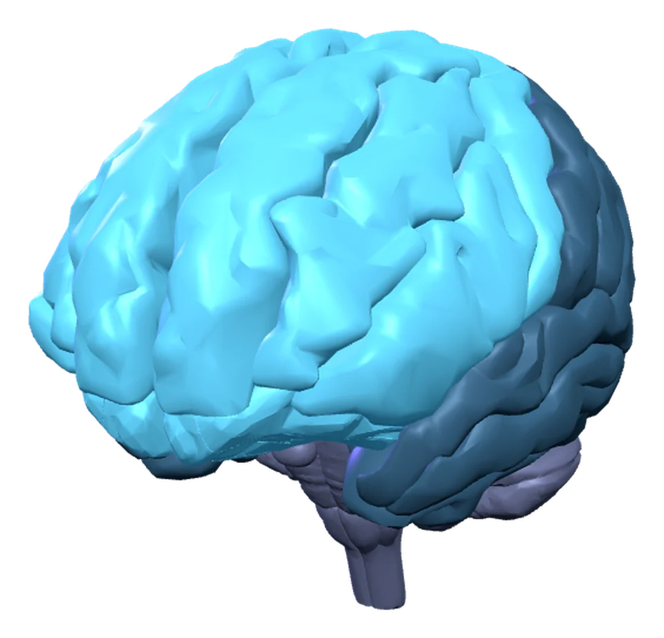
Choose a name to turn and highlight. Deep structures open a cutaway. Zoom with + / −; drag to inspect freely, then turn Drag Rotation Off to scroll.
Launch the lab to explore the model.
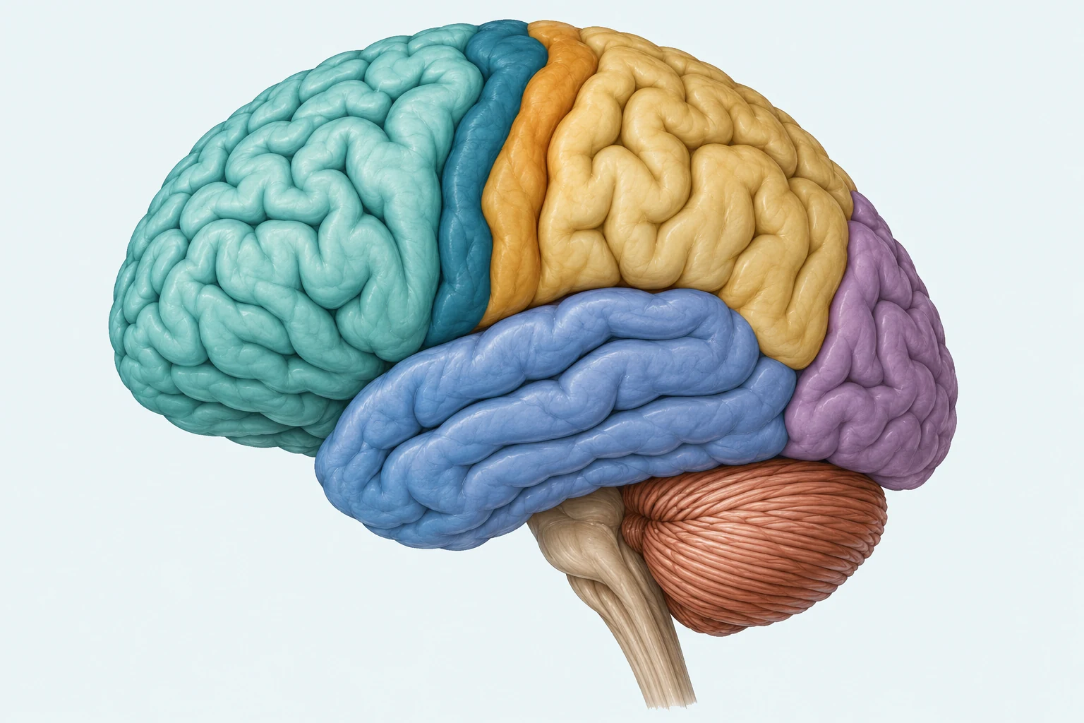
Match each numbered leader endpoint with its real location. The dark teal motor strip belongs to the posterior frontal lobe; the ochre somatosensory strip belongs to the anterior parietal lobe. Region coloring supports location, while the key supplies each functional distinction without relying on color.
- 1. Frontal lobe
- Front/left region: planning, language and voluntary movement.
- 2. Parietal lobe
- Upper rear region: touch processing and sensory integration.
- 3. Temporal lobe
- Lower lateral region: hearing and language processing.
- 4. Occipital lobe
- Posterior/right region: visual processing.
- 5. Motor strip
- Dark teal strip on the posterior frontal side of the central sulcus; helps direct skeletal movement.
- 6. Somatosensory strip
- Ochre strip on the anterior parietal side of the central sulcus; processes bodily touch.
- 7. Cerebellum
- Finely ridged structure beneath the posterior cerebrum: coordination and balance.
- 8. Brainstem
- Descending connection anterior to the cerebellum: basic bodily regulation.
Complete Structure Reference
- Brain stem
- Function: Supports basic bodily regulation and carries signals between brain and body.
Location: Midbrain, pons and medulla between the cerebrum and spinal cord, anterior to the cerebellum.
Application: Maintaining breathing while asleep relies on regulatory circuits rather than conscious rehearsal.Named anatomical surfaces from BodyParts3D 4.3. Simplified teaching geometry and lobe group boundaries; no neural activity is measured.
- Medulla
- Function: Helps regulate breathing and heart rate.
Location: Lower brain stem, below the pons and continuous with the spinal cord.
Application: An injury that disrupts medullary regulation can threaten vital functions even when remembered facts are not the main difficulty.Named anatomical surfaces from BodyParts3D 4.3. Simplified teaching geometry and lobe group boundaries; no neural activity is measured.
- Reticular activating system
- Function: Helps regulate alertness and wakefulness; related brainstem circuits contribute to eye and movement functions.
Location: Brainstem-centered ascending arousal circuitry interacting with deeper and cortical regions.
Application: Becoming alert to an important sound depends on interacting arousal circuits rather than one tiny attention switch.Schematic ascending-arousal connections between validated brainstem, thalamic and hypothalamic landmarks. No separately segmented RAS or exact tract route is supplied.
- Brain’s reward center
- Function: Contributes to motivation and learning from rewarding outcomes, alongside cognition and emotion.
Location: Distributed subcortical and cortical circuitry, rather than one bounded pleasure organ.
Application: A student becoming more likely to repeat a rewarded strategy illustrates learning from outcomes, not a scan proving a single reward spot activated.A sourced subset of participating orbitofrontal/frontal, amygdalar and substantia-nigra surfaces. Schematic connections are not a scan or one exclusive reward organ. VTA and nucleus accumbens contours are not available in this selected atlas.
- Cerebellum
- Function: Contributes to balance, movement coordination and some procedural learning.
Location: Lower rear of the brain, posterior to the brain stem.
Application: Smoothly timing a practiced sequence differs from simply deciding to start moving.Named anatomical surfaces from BodyParts3D 4.3. Simplified teaching geometry and lobe group boundaries; no neural activity is measured.
- Cerebral cortex
- Function: Supports connected sensory, motor and higher mental processing.
Location: Folded gray-matter outer layer of the cerebral hemispheres.
Application: Identifying a scene and organizing a response depend on communicating cortical regions, not one isolated all-purpose spot.Named anatomical surfaces from BodyParts3D 4.3. Simplified teaching geometry and lobe group boundaries; no neural activity is measured.
- Left cerebral hemisphere
- Function: Contains all four major lobes; language is typically more strongly left-associated and many pathways are contralateral.
Location: Anatomical left half of the cerebrum, separated from the right by the longitudinal fissure.
Application: With fixation, information flashed to the right visual field initially reaches the left hemisphere.Named anatomical surfaces from BodyParts3D 4.3. Simplified teaching geometry and lobe group boundaries; no neural activity is measured.
- Right cerebral hemisphere
- Function: Contains the same lobes and participates in connected processing; many sensory and motor pathways are contralateral.
Location: Anatomical right half of the cerebrum across the longitudinal fissure.
Application: A classic split-brain task can show left-hand selection of an object presented in the left visual field despite naming difficulty.Named anatomical surfaces from BodyParts3D 4.3. Simplified teaching geometry and lobe group boundaries; no neural activity is measured.
- Frontal lobes
- Function: Contributes to planning, language and voluntary movement.
Location: Anterior cortex, in front of the central sulcus and above the lateral sulcus.
Application: Organizing a sequence of actions uses frontal functions while sensory regions provide information about the situation.Named anatomical surfaces from BodyParts3D 4.3. Simplified teaching geometry and lobe group boundaries; no neural activity is measured.
- Parietal lobes
- Function: Contributes to organizing sensory information and bodily touch processing.
Location: Upper posterior cortex behind the central sulcus; posterior lateral boundary is partly conventional.
Application: Combining touch and position information can help locate an object relative to the body.Named anatomical surfaces from BodyParts3D 4.3. Simplified teaching geometry and lobe group boundaries; no neural activity is measured.
- Temporal lobes
- Function: Contributes to hearing and language processing.
Location: Lateral cortex below the lateral sulcus on both sides.
Application: Understanding a spoken instruction uses auditory and language networks that include temporal regions.Named anatomical surfaces from BodyParts3D 4.3. Simplified teaching geometry and lobe group boundaries; no neural activity is measured.
- Occipital lobes
- Function: Strongly involved in visual processing.
Location: Posterior cerebral cortex behind the parietal/temporal regions.
Application: Recognizing features of a displayed picture requires visual processing before a verbal explanation is produced.Named anatomical surfaces from BodyParts3D 4.3. Simplified teaching geometry and lobe group boundaries; no neural activity is measured.
- Prefrontal cortex
- Function: Supports planning, response inhibition and organizing goal-directed behavior.
Location: Anterior frontal cortex, ahead of premotor and primary motor regions.
Application: Delaying an impulsive response to follow a multistep plan illustrates executive functions.Approximate anterior frontal surface overlay, excluding the precentral motor gyri; no exact functional boundary is claimed.
- Motor cortex
- Function: Helps direct voluntary skeletal movement, largely on the opposite side of the body.
Location: Primary teaching focus: precentral gyri immediately anterior to the central sulcus.
Application: Reaching with the right hand largely involves motor output from the left hemisphere.Bilateral precentral gyri are gross anatomical anchors; this is not a fine map of every motor representation.
- Somatosensory cortex
- Function: Receives and processes bodily touch information.
Location: Primary teaching focus: postcentral gyri immediately posterior to the central sulcus.
Application: Identifying where a light touch occurred involves sensory processing rather than issuing a muscle command.Bilateral postcentral gyri are gross anatomical anchors; this is not a fine map of every body representation.
- Association areas
- Function: Integrate information and support connected higher functions.
Location: Distributed anterior, posterior and temporal cortical association regions.
Application: Recognizing an object and using its meaning to plan an action links different kinds of information.Representative distributed cortical surfaces, not one association organ and not an exhaustive network.
- Broca’s area
- Function: Contributes to producing language within a wider language network.
Location: Typical left caudal inferior frontal gyrus, including opercular and triangular regions.
Application: A person with disrupted language production may know what they intend to say yet struggle with fluent expression.Schematic location anchor on part of the left inferior frontal gyrus; broader than a precise functional or cytoarchitectonic boundary. Typical left specialization varies between people.
- Wernicke’s area
- Function: Contributes to understanding language within a wider language network.
Location: Typical left posterior temporal/temporoparietal language region.
Application: Fluent-sounding speech can coexist with difficulty understanding an instruction after disruption of language networks.Schematic location anchor on part of the posterior left superior temporal gyrus; the language-comprehension network extends beyond this teaching patch.
- Corpus callosum
- Function: Enables substantial communication between the hemispheres.
Location: Deep commissural white matter connecting the cerebral hemispheres.
Application: Severing this pathway can limit transfer needed to name information initially processed on the opposite side.Named anatomical surfaces from BodyParts3D 4.3. Simplified teaching geometry and lobe group boundaries; no neural activity is measured.
- Limbic system
- Function: Links memory, emotional significance and regulation through interacting structures.
Location: Connected cortical and deeper structures; membership varies across scientific accounts.
Application: An emotionally important event can involve both memory formation and bodily responses across a network.Course grouping of separate validated structures, with variable anatomical membership; not one enclosing organ.
- Thalamus
- Function: Routes most sensory information toward cortical processing; olfaction bypasses the initial relay.
Location: Paired deep nuclei on either side of the third ventricle, above the hypothalamus.
Application: A touch signal can be routed toward appropriate cortical processing rather than directly becoming a conscious conclusion at the receptor.Named anatomical surfaces from BodyParts3D 4.3. Simplified teaching geometry and lobe group boundaries; no neural activity is measured.
- Hypothalamus
- Function: Coordinates internal balance, bodily needs and endocrine regulation.
Location: Below and somewhat anterior to the thalamus, bordering the lower third ventricle.
Application: Temperature regulation or responding to hunger involves homeostatic coordination across neural and endocrine systems.Named anatomical surfaces from BodyParts3D 4.3. Simplified teaching geometry and lobe group boundaries; no neural activity is measured.
- Pituitary gland
- Function: Releases hormones under hypothalamic influence, linking neural regulation and endocrine functions.
Location: Below the hypothalamus, connected by the infundibular stalk at the brain’s base.
Application: Signals from hypothalamic control can influence hormone release and subsequent endocrine responses.Named anatomical surfaces from BodyParts3D 4.3. Simplified teaching geometry and lobe group boundaries; no neural activity is measured.
- Hippocampus
- Function: Important for forming new explicit memories of facts and events.
Location: Bilateral medial temporal structures, posterior to the amygdalae.
Application: Remembering a newly met person later illustrates memory formation, rather than storing all memories in one permanent cabinet.Named anatomical surfaces from BodyParts3D 4.3. Simplified teaching geometry and lobe group boundaries; no neural activity is measured.
- Amygdala
- Function: Participates in emotional significance and threat-related learning.
Location: Bilateral anterior medial-temporal nuclei, anterior to the hippocampi.
Application: A cue linked with an important outcome can later acquire emotional significance through interacting learning circuits.Named anatomical surfaces from BodyParts3D 4.3. Simplified teaching geometry and lobe group boundaries; no neural activity is measured.
Deeper Structures Connect Sensation, Regulation, Memory, and Emotion
The CED groups several structures under a limbic-system heading. Treat that grouping as a course organization of connected functions, not as a claim that every listed structure is cortical or that scientists draw exactly the same limbic boundary. These systems interact with the cortex and with one another.
The thalamus routes most sensory information toward appropriate cortical regions. Smell is the important exception to this initial relay. The hypothalamus helps regulate bodily needs and internal balance, including hunger, temperature, and aspects of endocrine control. It communicates with the pituitary gland, whose hormone releases influence other endocrine functions. This is the course’s permitted gland connection; a full gland-by-gland endocrine chart is unnecessary.
The hippocampus is important for forming new explicit memories, such as remembering a recent event or newly learned information. It is not a storage cabinet for every memory you will ever have. The amygdala participates in processing emotionally important information, including threat-related learning. It does not single-handedly produce all emotions. During an emotionally significant experience, memory and emotional processing can interact through these connected systems.
- Thalamus
- Sensory relay to cortex for most senses; olfaction bypasses this initial relay.
- Hypothalamus
- Regulation of internal bodily needs and endocrine coordination.
- Pituitary gland
- Hormone release under hypothalamic influence; connects neural regulation with endocrine functions.
- Hippocampus
- Formation of new explicit memories.
- Amygdala
- Processing of emotional significance, including threat-related learning.
- Corpus callosum
- Communication between the two cerebral hemispheres.
Language Specialization and the Logic of Split-Brain Tasks
In split-brain research, researchers study people whose corpus callosum has been severed, often as a treatment for severe epilepsy. Reduced communication between the hemispheres reveals aspects of lateralization, meaning that some functions are more strongly associated with one hemisphere. It does not establish that people have a logical-left or creative-right personality.
Language functions are usually more strongly associated with the left hemisphere. Broca’s area contributes to speech production, and Wernicke’s area contributes to language comprehension. Damage to language networks can cause aphasia, an impairment of language. Difficulty producing fluent speech differs from difficulty understanding language; neither should be confused with a general loss of intelligence.
Contralateral organization means that some information and control relate to the opposite side. With central fixation, a brief image in the left visual field is initially processed in the right hemisphere; a brief image in the right visual field is initially processed in the left hemisphere. A visual field is a region of space seen by both eyes, not the same thing as one eye. Most motor control of the left hand comes from the right hemisphere, and of the right hand from the left hemisphere.
In a classic task pattern, a split-brain participant might not verbally name an object flashed to the left visual field, because that information cannot readily reach the left hemisphere’s language system. The person may nevertheless choose the object with the left hand, which is controlled mainly by the right hemisphere. The object was processed; the difficulty is transferring information to the system needed for the requested response. Individual patterns vary, so present this as a demonstration of the task’s logic rather than a universal outcome.
Follow each brief visual-field presentation through the initially processing hemisphere to the response channel available in this classic task pattern.
Left Visual Field
Maintain central fixation · fields of view, not separate eyes
- Brief spoon image
- Right hemisphere
- Left-hand selection

Can select the spoon with the left hand even when the information cannot readily reach the left-language system for verbal naming.
Right Visual Field
Maintain central fixation · fields of view, not separate eyes
- Brief spoon image
- Left hemisphere
- Verbal naming

The left hemisphere’s usual language access supports saying “spoon.”
Between hemispheres: corpus callosum severed → information transfer restricted.
Plasticity and Evidence About Brain Function
Brain plasticity is the capacity to change connections and patterns of functioning through development, experience, and sometimes recovery after injury. Practice can alter networks, and some functions can be partly supported by other regions after damage. Plasticity is constrained: compensation is not a guarantee that any damaged area can be completely replaced.
An EEG (electroencephalogram) records electrical activity using scalp electrodes. It is especially useful for changes over time, including sleep-stage patterns. fMRI (functional magnetic resonance imaging) uses changes related to blood oxygenation as an indirect indicator of activity and helps locate regions associated with a task. A colorful activation map does not establish that one region alone causes a behavior, and blood-oxygen signals are not direct pictures of individual neurons firing.
Case studies examine an individual in depth and can reveal valuable patterns after unusual injuries; they are limited in generalization. Lesioning damages or removes tissue and can help establish how disrupted structures affect functioning, but planned harmful lesions in healthy humans are not an ethical experimental method. Researchers may study naturally occurring injuries, clinically necessary surgery, or ethically reviewed animal work. Method, ethical safeguards, and converging evidence determine what a study can support.
| Evidence source | What it reveals | What to avoid concluding |
|---|---|---|
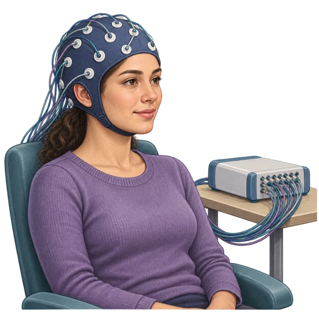 EEG EEG | Changes in recorded electrical activity over time | What to avoid concludingIt pinpoints every active neuron. |
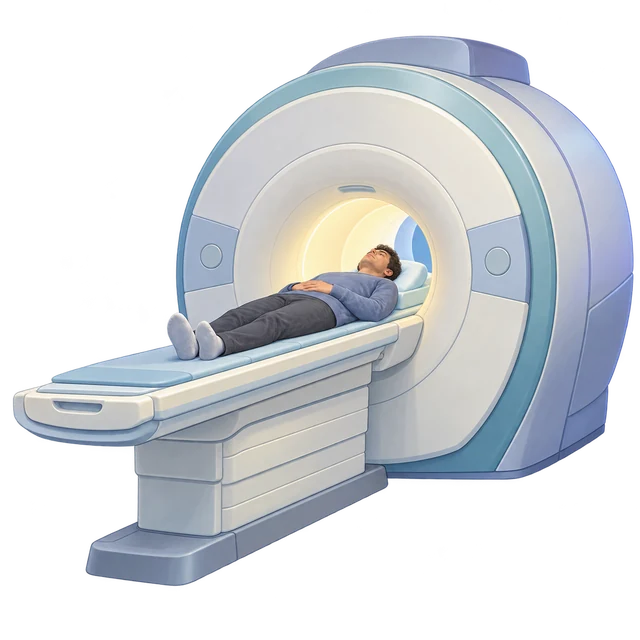 fMRI fMRI | Blood-oxygen changes associated with a task | What to avoid concludingActivation by itself proves a region is the sole cause. |
 Case study Case study | A detailed pattern in an individual | What to avoid concludingOne case necessarily describes everyone. |
| Lesion or surgery evidence | Consequences of disrupted tissue | What to avoid concludingResearch benefit makes harmful human procedures acceptable. |
Compare electrical timing, blood-oxygen-related activity, an individual case and the effects of damage or intervention. Each evidence source supports a different inference; use the caution column to limit the conclusion.
Sleep
Sleep
Sleep is an active, changing state of consciousness. Its timing, stages, and disruptions help explain how biology affects attention, memory, and everyday performance.
CED printed pp. 41–42Awareness Changes; Circadian Timing Organizes the Day
Consciousness is awareness of thoughts, feelings, behavior, and internal or external events. Its level and contents vary. Wakefulness and sleep are different states of consciousness; sleep is not simply an inactive brain or the complete absence of mental processes.
A circadian rhythm is an approximately 24-hour biological cycle. The sleep/wake cycle is one example. Internal timing interacts with environmental cues, particularly light, and with schedules. Melatonin contributes to signaling biological night. The rhythm influences when sleep and alertness are likely, even though obligations can make a person stay awake at a biologically inconvenient time.
Jet lag follows a rapid shift across time zones when internal timing has not yet aligned with local day and night. Shift work can require activity during biological night and sleep during biological day. Social expectations and work or school schedules therefore interact with biological timing. A person’s difficulty maintaining attention on an overnight shift need not indicate laziness; the circumstances may be misaligned with the sleep/wake rhythm.
A sleep/wake rhythm runs on an approximately 24-hour cycle. Light and schedules interact with internal timing; the timing of a sleep stage is a different question.

Local Light + Schedules
Day/night cues and school or work schedules shape the timing context.

Internal Biological Timing
The approximately 24-hour rhythm influences likely sleep and alertness.
Environmental cues ↔ internal timing: alignment can change with jet lag or shift work.
Read Sleep as a Changing Sequence of States
An EEG helps distinguish stages by patterns of brain electrical activity. NREM (non-rapid eye movement) sleep has three stages. N1 is the transition into light sleep, with slower activity than alert wakefulness; hypnagogic sensations, such as a falling sensation, may occur as a person enters it. N2 is characterized by bursts called sleep spindles and large brief events called K-complexes. N3 is deep, slow-wave sleep with prominent slow, high-amplitude activity.
REM (rapid eye movement) sleep involves relatively active, low-amplitude mixed-frequency brain activity resembling wakefulness, while most voluntary skeletal muscles are strongly relaxed. This combination explains the term paradoxical sleep: an active-looking EEG accompanies marked muscle relaxation. Eye movement and essential bodily functions continue; the entire body is not motionless. Vivid dreaming is especially associated with REM, although dreams can also occur in NREM.
The stages recur through the night, rather than progressing once from N1 to N3 to REM and stopping. Deep N3 sleep is more concentrated early in the night, while REM periods generally become longer later. The CED describes NREM decreasing and REM increasing as the cycle progresses; read this as a changing pattern across repeated nighttime cycles, not a claim that every NREM stage must shorten on every pass.
After REM deprivation, REM rebound can occur: subsequent sleep contains increased REM relative to the person’s previous pattern. Rebound is a compensatory pattern, not evidence that a person can permanently eliminate a needed stage.
Sleep Cycle
Explorer
Follow the Night. Read the Pattern.
One authored night with five varied cycles, not a personal record or a sleep-duration recommendation.

State
Cues are qualitative. A facial expression cannot identify a sleep stage.
Low-amplitude, fast mixed activity in this alert-wake example. Relaxed wakefulness can look different.
Before Sleep / Brief Awakening
Wakefulness is a distinct state. This authored night includes a short settling period and brief awakenings between some cycles.
The waveform is constructed, not recorded EEG. Its short window is separate from the hours-long timeline.
Sleep Stages: Complete Reference
The original six-hour static illustration is retained below. The interactive eight-hour night is a separate authored example of the same early-NREM/later-REM pattern.
- Stage Axis
REM, N1, N2, and N3 are categories, not numeric measurements.
- Time Axis
0–6 elapsed hours after sleep begins.
- Read the Line
N3 is concentrated early; later REM periods are longer.
- N1 · Transition
Light sleep; hypnagogic sensations may occur.
- N2 · Established Sleep
EEG shows spindles and K-complexes.
- N3 · Slow-Wave
Prominent slow, high-amplitude EEG activity.
- REM · Paradoxical
Wake-like activity plus marked voluntary-muscle relaxation.
The Interactive Night’s Dataset
| Cycle | Stage | Elapsed Time | Minutes |
|---|---|---|---|
| Settling | Awake | 0:00–0:05 | 5 |
| 1 | NREM 1 | 0:05–0:10 | 5 |
| 1 | NREM 2 | 0:10–0:30 | 20 |
| 1 | NREM 3 | 0:30–1:05 | 35 |
| 1 | NREM 2 | 1:05–1:22 | 17 |
| 1 | REM | 1:22–1:35 | 13 |
| 2 | NREM 1 | 1:35–1:39 | 4 |
| 2 | NREM 2 | 1:39–2:04 | 25 |
| 2 | NREM 3 | 2:04–2:29 | 25 |
| 2 | NREM 2 | 2:29–2:49 | 20 |
| 2 | REM | 2:49–3:08 | 19 |
| 2 | Awake | 3:08–3:10 | 2 |
| 3 | NREM 1 | 3:10–3:13 | 3 |
| 3 | NREM 2 | 3:13–3:44 | 31 |
| 3 | NREM 3 | 3:44–3:56 | 12 |
| 3 | NREM 2 | 3:56–4:17 | 21 |
| 3 | REM | 4:17–4:42 | 25 |
| 3 | Awake | 4:42–4:45 | 3 |
| 4 | NREM 1 | 4:45–4:48 | 3 |
| 4 | NREM 2 | 4:48–5:50 | 62 |
| 4 | REM | 5:50–6:23 | 33 |
| 4 | Awake | 6:23–6:25 | 2 |
| 5 | NREM 1 | 6:25–6:28 | 3 |
| 5 | NREM 2 | 6:28–7:20 | 52 |
| 5 | REM | 7:20–7:58 | 38 |
| 5 | Awake | 7:58–8:00 | 2 |
Awake · Before Sleep / Brief Awakening
Wakefulness is a distinct state. This authored night includes a short settling period and brief awakenings between some cycles.
Low-amplitude, fast mixed activity in this alert-wake example. Relaxed wakefulness can look different.
REM · Active Brain, Relaxed Muscles
Wake-like brain activity accompanies marked relaxation of most voluntary skeletal muscles. Eye movements and essential functions continue. Vivid dreaming is especially associated with REM, but dreams also occur in NREM.
Low-amplitude mixed-frequency activity resembles the awake example. The muscle-state cue makes the REM contrast clear.
NREM 1 · The Transition into Sleep
Light sleep begins. Hypnagogic sensations, such as a feeling of falling, may occur during this transition; they are not required on every pass.
Slower theta-like activity, with a fading alpha-like component, distinguishes this transition from alert wakefulness.
NREM 2 · Spindles and K-Complexes
Established NREM sleep has characteristic EEG events: a sleep spindle is a brief faster burst; a K-complex is a large, brief event.
Gold Region: Spindle · Blue Region: K-Complex
A spindle around 1 second and a biphasic K-complex around 2.4 seconds are highlighted on a slower background.
NREM 3 · Deep, Slow-Wave Sleep
Prominent slow-wave activity distinguishes NREM 3. This example concentrates it in the first three cycles, especially the first.
Large, slow delta-like waves dominate this schematic. The height is relative, not calibrated EEG voltage.
Early / Late Comparison
Cycle 1 · 90 Minutes
0:05–1:35 elapsed
| Stage | Minutes |
|---|---|
| Awake | 0 |
| REM | 13 |
| NREM 1 | 5 |
| NREM 2 | 37 |
| NREM 3 | 35 |
Cycle 5 · 95 Minutes
6:25–8:00 elapsed
| Stage | Minutes |
|---|---|
| Awake | 2 |
| REM | 38 |
| NREM 1 | 3 |
| NREM 2 | 52 |
| NREM 3 | 0 |
In this same night, NREM 3 falls from 35 minutes in Cycle 1 to 0 in Cycle 5; REM rises from 13 to 38 minutes. Stage boundaries and durations vary in real nights. Color bands use the timeline’s stage order and colors.
Sources and Example Limits
AP scope: current CED 1.5.A.3, .i and .ii, printed p. 41 / PDF p. 48; October 2025 corrections use “hypnagogic.” NHLBI supports repeated cycles, early deep sleep and later REM. OpenStax: sleep stages supports the schematic distinctions.
Stage boundaries, waves and qualitative cues are authored illustrations. They are not Mr. Steadman’s measurements, diagnostic criteria or a universal timetable. The eight-hour duration sets the display window. Playback compresses time while allowing at least 2.5 seconds per segment. Existing circadian, dream-theory, sleep-purpose, REM-rebound and disorder explanations remain in the article.
Dreaming, Memory Consolidation, and Restoration
Activation-synthesis theory proposes that the brain constructs a meaningful dream from internally generated activity during sleep. It explains the story-like experience as an interpretation of that activity, rather than treating every dream detail as a hidden message.
Consolidation theory connects sleep and dreaming with processing and strengthening information from waking experience. Memory consolidation is the process by which new memories become more stable and organized. Incorporating experiences into dreams is compatible with this account, but one dream cannot prove exactly how a memory was consolidated.
Restoration theory proposes that sleep helps replenish resources and supports repair and recovery after waking activity. Restoration and memory consolidation address different functions that can both contribute to why organisms sleep. They need not be mutually exclusive answers. The psychoanalytic theory of dreams is outside this topic’s AP exam scope.
| Account | Central proposal | Useful distinction |
|---|---|---|
| Activation-synthesis | The brain interprets internally generated activity into dream experience. | Explains construction of a dream. |
| Consolidation | Sleep supports stabilization and organization of memories. | Connects sleep with information processing. |
| Restoration | Sleep helps replenish resources and support recovery. | Emphasizes maintenance and recovery functions. |
The proposals explain different questions about dreaming and sleep; a plausible explanation still needs evidence.
Different Disruptions, Different Waking Consequences
Disrupted sleep can impair attention, memory, response speed, mood regulation, and physical performance. Distinguish disorders by the process disrupted, rather than labeling every tired person with insomnia. The course specifies the five disorders below; the descriptions are instructional distinctions, not a basis for self-diagnosis.
Treatment appropriate to the disorder and a regular sleep schedule can improve waking functioning and well-being. The explanatory connection matters: repeated awakenings can reduce restorative sleep, which can impair daytime attention even when time in bed looks adequate. A regular schedule helps align timing, but it is not a cure for every disorder.
- Insomnia
- Persistent difficulty falling asleep, staying asleep, or obtaining satisfactory sleep, with resulting daytime effects. It differs from choosing not to allow enough time for sleep.
- Narcolepsy
- A disorder involving marked daytime sleepiness and recurrent episodes of sleep; REM-related phenomena can intrude into waking life. It is not ordinary tiredness after a late night.
- REM sleep behavior disorder
- Normal REM muscle inhibition is disrupted, allowing a person to physically act out dream-related movements.
- Sleep apnea
- Repeated interruptions or reductions in breathing during sleep that disrupt sleep and can cause daytime sleepiness.
- Somnambulism
- Sleepwalking, usually associated with deep NREM sleep. Contrast it with dream enactment in REM sleep behavior disorder.
Sensation
Sensation
Sensory systems convert physical or chemical stimulation into neural messages. Connect detection, transduction, and bodily or brain processing; then distinguish what different systems contribute.
CED printed pp. 43–46Detection Is Not the Same as Interpretation
Sensation is detecting stimulation and converting it into signals the nervous system can use. Transduction is the conversion from physical or chemical stimulation into neural signals. Perception is organizing and interpreting sensory information. Light reaching receptors, receptor transduction, and recognizing a familiar face are connected events, but the last is perception rather than simple detection.
The absolute threshold is the minimum intensity detected on at least 50% of trials under specified conditions. It is a probability-based boundary, not a promise that every stimulus at that intensity is detected. A threshold study must state what stimulus varies, how detection is measured, and what conditions are held constant.
The just-noticeable difference (JND), or difference threshold, concerns the smallest detectable change between stimuli. Weber’s law proposes that the change needed for detection is approximately a constant proportion of the starting intensity, within a relevant range. A 2-gram difference can matter when comparing very light weights but be imperceptible when added to a heavy object. Absolute threshold asks whether stimulation is present; JND asks whether it changed.
Sensory adaptation is reduced sensitivity during sustained, unchanging stimulation, such as noticing a steady odor less after entering a room. It does not mean that the odor physically disappeared. Distinguish adaptation from intentionally shifting attention and from habituation, a learned reduction in response discussed in Unit 3.
Sensory Threshold
Workbench
Two Questions About What We Notice.
One Tone, Ten Attempts
Absolute threshold is the minimum stimulus intensity detected on at least half the attempts, under specified conditions.
In this authored example, imagine one listener given a tone ten times at each intensity level. One trial is one presentation of the tone—one opportunity to detect it.
The tone’s intensity stays the same within each set of ten. Choosing another level changes the relative sound intensity: Level 1 is weakest; Level 5 is strongest.
“Half the time” means half the detection attempts (50%). It does not mean half volume, half the people, or part of a sound.
Choose a Tone Intensity Level
Heard: 5 of 10
Not Heard: 5 of 10
Each marker represents one attempt. ✓ Heard: the listener detected the tone. – Not Heard: the listener did not detect it. These are grouped counts, not an order of trials or ten people.
5 of 10 is half the attempts (50%). This is the lowest tested level detected on at least half the attempts. It meets the absolute-threshold criterion in this example.
These invented counts illustrate the criterion; no experiment or hearing test is being conducted. Five tested levels do not locate an exact continuous biological threshold. No audio is needed.
See the Same Results on a Graph
Vertical: Attempts Heard (%) · Dashed Line: 50%
Outlined Bar: Level 3 · 50% Heard
Show the Full Example Table
| Level | Heard | Not Heard | Heard (%) |
|---|---|---|---|
| 1 | 1 / 10 | 9 / 10 | 10% |
| 2 | 3 / 10 | 7 / 10 | 30% |
| 3 | 5 / 10 | 5 / 10 | 50% |
| 4 | 8 / 10 | 2 / 10 | 80% |
| 5 | 10 / 10 | 0 / 10 | 100% |
Absolute threshold: detecting a stimulus, as with the tone. JND: detecting a difference, as between the two weights. Weber’s law: relating the needed difference to the starting amount.
These sensory thresholds differ from Neuron Lab’s membrane-firing threshold.
Thresholds: Complete Reference
Absolute threshold is the minimum intensity detected on at least 50% of trials under specified conditions. It is a probability-based boundary, not a guarantee of detecting every presentation or a bodily switch.
The authored tone example uses one listener and ten attempts at each relative intensity. Each trial is one tone presentation; intensity stays fixed within a set and changes between levels. “Heard” means detected; “Not Heard” means not detected. The counts below are grouped, not a temporal record. Half the time refers to attempts, not volume or people.
- Intensity Axis
Tone levels 1–5, in arbitrary units.
- Detection Axis
Percent of ten trials where the tone was reported heard.
- Detection Pattern
Levels 1–5: 10%, 30%, 50%, 80%, 100%. Level 3 reaches the 50% threshold.
Presence: absolute threshold · Change: JND · Constant input: adaptation
| Level | Heard | Not Heard | Heard (%) |
|---|---|---|---|
| 1 | 1 / 10 | 9 / 10 | 10% |
| 2 | 3 / 10 | 7 / 10 | 30% |
| 3 | 5 / 10 | 5 / 10 | 50% |
| 4 | 8 / 10 | 2 / 10 | 80% |
| 5 | 10 / 10 | 0 / 10 | 100% |
Level 3 is the lowest tested level detected on at least half the attempts (5 of 10). Level 1 and Level 2 include some detections but fall below that criterion. Level 4 and Level 5 are easier to detect in these results, but Level 3 already qualifies. These five relative teaching levels are not calibrated decibels or an exact continuous biological threshold.
JND and Weber’s Law
The just-noticeable difference (JND), or difference threshold, concerns the smallest detectable change between stimuli under specified conditions. Weber’s law proposes that the change needed for detection is approximately a constant proportion of the starting intensity, within a relevant range.
In the preserved model, the original weight is 100 g and adding 5 g gives a comparison weight of 105 g. The JND is the 5 g difference, not the 105 g total. No addition gives a 0 g difference; adding 2 g is below this model’s 5 g JND.
A: start at 100 g and add 5 g. B: double only the start to 200 g and keep the 5 g addition; the 205 g total now has a smaller relative difference, below the model’s 10 g JND. C: keep the 200 g start and increase the addition to 10 g; the 210 g total reaches the same proportional difference criterion.
- A · Start 100 g · Add 5 g
Comparison Total: 105 g
5 / 100 = 5%
- B · Start 200 g · Add 5 g
Comparison Total: 205 g
5 / 200 = 2.5%
- C · Start 200 g · Add 10 g
Comparison Total: 210 g
10 / 200 = 5%
5 / 100 = 0.05 · 0.05 × 200 = 10 g
The 5% fraction is an assumption for this example and range, not a universal rule or a guarantee of what a person notices. The displayed weights are not a test of detecting weight. Sensory adaptation and the other sensation explanations remain in the complete article. No hearing or other personal sensory test is conducted. These sensory thresholds differ from Neuron Lab’s membrane-firing threshold.
Sources and Example Limits
Current CED 1.6.A.1–1.6.A.2, printed p. 43 / PDF p. 50, defines the curricular scope. OpenStax: sensation and perception supports the threshold/proportion distinction. The unchanged article and static figure supply the counts and mass example. Trial setup and guide are explicitly authored context. The five levels and 5% fraction are teaching choices, not official numerical requirements or individual measurements. Conditions affect detection.
Senses Can Combine Without Becoming Interchangeable
Sensory interaction occurs when information from one sense influences the experience produced with another. Food flavor integrates taste, smell, texture, and temperature. A blocked nose can change flavor even while taste receptors still detect sweetness or saltiness.
Synesthesia is a consistent experience in which stimulation in one sensory or cognitive system is accompanied by an experience in another, such as sounds eliciting color experiences. This differs from ordinary sensory interaction: hearing a note and consistently experiencing a color is not the same as smell contributing information about food. It also differs from a metaphor such as calling a sound bright.
Light Must Be Focused and Transduced
The retina is the light-sensitive surface at the back of the eye. Photoreceptors transduce light, and retinal circuits carry information toward the brain through the optic nerve. The blind spot is the retinal location where the optic nerve exits; it lacks photoreceptors. Perception usually feels continuous because the brain combines available information and fills gaps rather than delivering a photograph of each receptor’s input.
The lens changes shape during accommodation to help focus light onto the retina. This is an optical focusing process; it is different from the schema-changing accommodation in cognition and development. Nearsightedness means distant objects are hard to focus sharply, commonly because light focuses in front of the retina. Farsightedness involves difficulty with near focus, commonly because light would focus behind the retina without sufficient compensation. Both illustrate a mismatch in optical focusing, rather than a failure of the occipital lobe to interpret an otherwise sharp image.
EYE LAB
Explore the Eye. Follow the Light. Bring It into Focus.
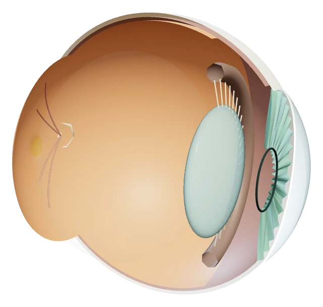
Choose a structure or select a visible surface. View buttons work without dragging. The cutaway opens the temporal side; the pupil is an opening.
Eye Lab: Complete 2D Reference
Explore a right eye, follow light into retinal transduction, compare optical focus and inspect receptor distribution. The model’s additional anatomy supports understanding; it is not an extra AP memorization list.
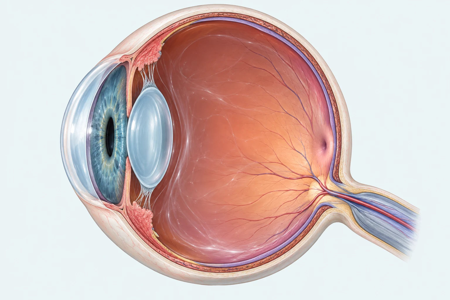
The lens changes shape during accommodation to focus light onto the retinal surface. Retinal cells transduce stimulation; the fovea supports detailed fixation. The optic nerve carries neural messages, while its separate retinal exit forms the blind spot.
- 1. Lens
- The translucent biconvex lens behind the iris changes shape to focus incoming light.
- 2. Retina
- The inner lining of the posterior eye wall contains cells that transduce light.
- 3. Fovea
- The posterior retinal depression supports cone-rich detailed fixation; it is not the nerve exit.
- 4. Blind spot
- The optic-disc exit at the lower posterior retina has no photoreceptors; vessels converge here.
- 5. Optic nerve
- The nerve exits the posterior wall and carries retinal signals toward the brain.
Solid ray: simplified incoming light direction through the lens toward the retina.
Explore the Eye
- Cornea
- Function: Bends entering light and protects the front of the eye.
Location: Transparent dome in front of the iris. Its curvature supplies much of the eye’s optical power. Supporting anatomy.
- Iris
- Function: Changes the pupil opening to regulate how much light enters.
Location: Colored diaphragm between the cornea and lens. In the modeled dim scene, its opening widens; the lens setting does not change. Supporting anatomy.
- Pupil
- Function: Provides the opening through which light enters the eye.
Location: The empty central opening in the iris, in front of the lens. It is an aperture, not a black piece of tissue. Supporting anatomy.
- Lens
- Function: Changes shape during accommodation to help focus light onto the retina.
Location: Clear, biconvex structure behind the iris. Near focus uses a rounder, thicker lens; distant focus uses a flatter lens. AP 1.6.B.2.
- Retina
- Function: Transduces light and begins processing visual information.
Location: Light-sensitive lining of the posterior eye wall. Photoreceptors, bipolar cells and ganglion cells form a simplified route through retinal circuits. AP 1.6.B.1 / B.3.
- Fovea
- Function: Supports fine detail and color during central fixation through its dense cone population.
Location: Small temporal-side depression in the posterior retina, distinct from the optic disc. Reading fine print benefits from cone-rich central vision. The foveal center lacks rods. AP 1.6.B.4.i.
- Optic Disc / Blind Spot
- Function: Provides the exit for ganglion-cell axons; it has no photoreceptors.
Location: Nasal to the fovea in the posterior right-eye retina. Its missing receptors create a blind spot in the temporal visual field. AP 1.6.B.1.
- Optic Nerve
- Function: Carries neural information from retinal ganglion cells toward the brain.
Location: Leaves the posterior eye at the optic disc, on the nasal side of the fovea. Neural signals travel here, not the optical retinal image. AP 1.6.B.1 / supporting pathway.
- Sclera
- Function: Forms a protective outer coat and supports the shape of the eye.
Location: Opaque outer wall surrounding most of the globe. The cutaway removes part of this wall to reveal the interior. Supporting anatomy.
- Ciliary Body and Zonules
- Function: Adjust lens shape by changing tension in the fibers that suspend it.
Location: Ring around the lens; zonular fibers connect it to the lens rim. For near accommodation, ciliary muscle contraction reduces zonular tension so the lens rounds. Supporting anatomy.
- Vitreous Chamber
- Function: Provides a clear medium through which light reaches the retina.
Location: Large cavity behind the lens, filled by vitreous gel in the living eye. The transparent cue identifies the space without obscuring the retinal lining. Supporting anatomy.
Follow the Light
- Light from an Object. The arrow has a tip and a base. Light from each feature travels toward the eye; the arrow itself stays outside.
- Cornea Bends Light. The transparent, curved cornea bends incoming light. It supplies much of the eye’s focusing power.
- Through the Pupil. Light passes through the opening in the iris. Changing its size controls light entry; this is distinct from accommodation.
- Lens Refines Focus. The lens works with the cornea to focus light on the retina. Rays from the arrow’s tip and base reach different retinal positions.
- Retinal Image and Transduction. The optical image is inverted on the retina. Photoreceptors convert light into changes in cellular signaling; a little photograph does not enter the nerve.
- Retinal Circuit Carries Information. Photoreceptors influence bipolar cells, which influence ganglion cells. Photoreceptor and bipolar responses are generally graded; ganglion-cell axons carry action potentials.
- Neural Information toward the Brain. Ganglion-cell axons leave through the optic disc and optic nerve. This purple cue represents neural information toward visual pathways, not a light beam or a traveling picture.
A generic retinal section, linked to the lining highlighted in the eye. The foveal pit displaces inner layers. Light travels toward outer receptor segments; neural influence travels back through retinal circuits. Cell sizes, spacing and connections are schematic.
Focus the Image
Automatic accommodation rounds the lens for near focus and flattens it for distant focus. Ciliary contraction reduces zonular tension for near accommodation. Iris/pupil adjustment controls light entry separately. A held lens can leave the image out of focus when object distance changes; that setting is not itself a refractive error.
Gold: optical rays. Dashed gold beyond the retina: geometric continuation, not light passing through the eye wall. The cornea and lens are combined into one effective optical plane in this calculation; object distance is compressed to fit the view.
Focus on the Retina
On the Retina. Retina distance: 2.85 units.
Myopic Example
In Front of the Retina. Retina distance: 3.35 units.
Hyperopic Example
Behind the Retina. Retina distance: 2.35 units.
These refractive examples hold the distant object and relaxed whole-eye power equal while changing retinal distance. Farsightedness does not guarantee clear distant vision. Dashed rays behind the retina are geometric continuations, not light leaving the back wall.
Retina Workshop
Schematic density pattern, not a count or sensitivity measurement. Cones also occur outside the fovea. Dim/bright selects a viewing condition; biological adaptation takes time.
The foveal center contains densely packed cones and lacks rods. Peripheral retina contains many rods and some cones. Rods support dim-light detection without color distinctions; cones support color and detail in sufficient light. Environment changes do not instantly complete biological adaptation.
Optional Blind-Spot Reference
The optic disc, nasal to the right-eye fovea, lacks photoreceptors and maps to the temporal visual field. For the optional demonstration, cover the left eye gently, fixate the left cross with the right eye, and briefly vary the separation of the dot to its right or a comfortable viewing distance. Both marks stay present. Not noticing an effect is possible; it is not an eye-health result. Skip or stop if uncomfortable.
The original rods/cones comparison, trichromatic theory, opponent processing, color-vision differences and visual-disorder explanations remain in the article below.
Sources and Model Assumptions
AP scope follows the current AP Psychology CED, Topic 1.6.B, printed pages 43–44, with October 2025 clarifications. The linked file has a Fall 2025 cover and a Fall 2024 inner title; no vision correction appears in the clarifications.
Anatomy and function: NEI: How Eyes Work, NEI: About the Eye, NEI: Refractive Errors, UTHealth: Eye and Retina and UTHealth: Visual Pathways.
The original procedural right-eye geometry uses anterior +X, superior +Y, temporal +Z; the optic disc is nasal to the fovea. Partial coats expose the interior. Colors, layer thickness, vessel detail, receptor spacing and cutaway are teaching choices, not individual clinical anatomy. The pupil is an actual opening; its invisible picking surface has no rendered color. Chamber and ciliary cues are simplified.
Focus uses a paraxial thin-lens approximation: 1/u + 1/v = P; image height = −v/u × object height. Arbitrary length units use retina distance 2.85, object range 6–30 and aperture half-height 0.30. The effective whole-eye power combines corneal and lens refraction. Accommodation varies P from 1/2.85 + 1/30 to 1/2.85 + 1/6. Automatic mode sets P = 1/2.85 + 1/u. Manual percent is a model interpolation, not measured muscle effort or a prescription. Blur radius is half the ray-bundle width at the retina, converted at 56 CSS pixels per model unit to a bounded preview blur. Retina distance uses a flat equivalent image plane at x = −1.90; the anatomical lining is curved, so off-axis endpoints can differ slightly from that surface. The corneal entry is schematic, with straight passage through the pupil; exact ray calculation starts at the effective plane represented at the lens.
Retinal slabs omit interneuron detail and are not exact foveal sections. Foveal/peripheral drawings show schematic density, not exact cell counts. Optical light and neural signaling have distinct cues. The pathway toward the brain is abbreviated. Model construction and provenance.
Receptor Types Explain Sensitivity, Detail, and Adaptation
Rods are especially useful in dim light and support sensitivity to light, shapes, and movement without color discrimination. They are more abundant away from the foveal center. When entering darkness, sensitivity changes as visual systems adapt; rods are especially important in dark adaptation. Adaptation also occurs when returning to brighter conditions.
Cones support color vision and fine detail in sufficient light. They are densely concentrated in the fovea, the central region used for detailed fixation. Cones also occur elsewhere in the retina, so foveal concentration does not mean that cones exist only there. A faint object may be easier to detect slightly away from direct fixation, while reading small print benefits from central cone-rich vision.
The three cone types respond most strongly to different, overlapping ranges of wavelengths: short, medium, and long. These are often called blue, green, and red cones. A cone is not a tiny blue, green, or red picture sensor; the brain uses their relative responses to encode color.
| Feature | Rods | Cones |
|---|---|---|
Main strength | Dim-light sensitivity; shapes and movement | Color and fine detail in brighter conditions |
Distribution | More prominent outside the foveal center | Densely concentrated in the fovea |
Color discrimination | Does not support color distinctions | Relative responses of three types support color vision |
Two Color Theories Describe Different Processing Stages
Trichromatic theory explains color coding through the combined activity of three cone types. Different patterns of short-, medium-, and long-wavelength responses support different color experiences. Opponent-process theory explains further processing in opponent channels: red versus green, blue versus yellow, and black versus white. Increasing one side of an opponent signal is associated with decreasing the other side.
The theories complement one another. Cones provide inputs; retinal ganglion cells and later pathways compare those inputs. After prolonged exposure to a color, adaptation within these systems can contribute to an afterimage in its opponent direction. Looking at an intense red region and then a neutral surface can produce a greenish afterimage. This does not mean that a new green object appeared on the surface.
Color vision deficiency involves altered color processing, often because a cone class functions differently or is absent, and sometimes because of other visual-pathway problems. Dichromatism involves two functioning cone classes rather than the typical three; monochromatism involves only one functional color channel or severe absence of ordinary cone-based color discrimination. Color deficiency is not necessarily seeing only black and white. The CED also references irregularities involving opponent ganglion-cell processing; distinguish receptor coding from later comparisons rather than treating every deficit as identical.
The two color theories explain different stages: receptor inputs first, then comparisons of those inputs in retinal and later pathways.
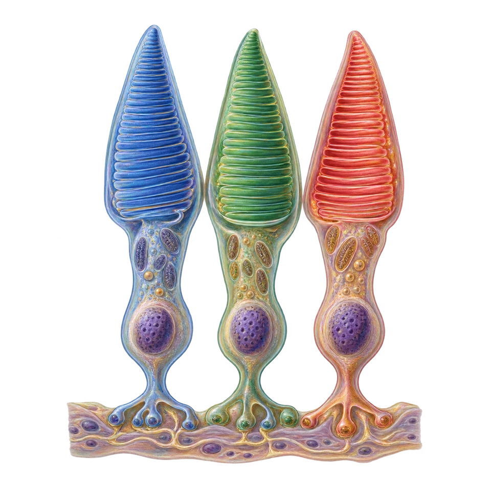 Stage 1
Three Receptor Inputs
Short-, medium-, and long-wavelength cones respond in overlapping ranges.
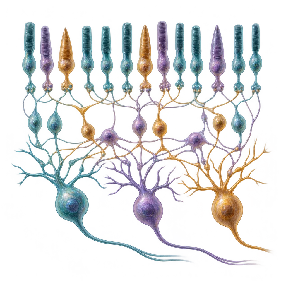 Stage 2
Opponent Comparisons
Retinal and later pathways compare signals: red/green, blue/yellow, black/white.
 Stage 3
Experience and Adaptation
Combined activity supports color perception; sustained exposure can produce an opponent-direction afterimage.
Detecting a Stimulus and Recognizing It Can Separate
Visual function depends on brain processing as well as healthy eyes. Damage in visual pathways, including occipital and connected regions, can disrupt aspects of vision. Prosopagnosia, or face blindness, involves difficulty recognizing faces even when other visual abilities are relatively intact. It is associated with specialized face-processing networks, including occipitotemporal areas, rather than being a generic synonym for all occipital damage.
In blindsight, a person with damage affecting primary visual processing may respond to some visual information in a region reported as unseen, without normal conscious visual awareness there. For instance, performance in locating a stimulus may exceed what random guessing would predict, despite the person saying they did not consciously see it. This illustrates a distinction between some information processing and conscious perception; it does not mean normal vision is fully preserved.
Pitch and Loudness Describe Different Wave Properties
Sound is a pattern of pressure changes produced by vibrating matter. Frequency, the number of cycles per second measured in hertz (Hz), is closely related to perceived pitch. Higher-frequency sound generally has higher pitch. In a given medium, shorter wavelength corresponds to higher frequency. The physical wave property and the subjective pitch experience are related but are not the same thing.
Amplitude, the size of pressure variation, is related to perceived loudness. Increasing amplitude generally makes a sound louder, without necessarily changing its frequency or pitch. Loudness also depends on frequency and the listener. Therefore, tall waves are not automatically high-pitched waves.
- Horizontal Axis
Time in milliseconds, 0–20 ms in each panel.
- Vertical Axis
Pressure deviation in arbitrary units, from −2 to +2.
- A · Reference
100 Hz, amplitude 1.
- B · Frequency Changes
200 Hz, amplitude 1: more cycles in the same time, higher pitch.
- C · Amplitude Changes
100 Hz, amplitude 2: taller wave, usually louder; pitch unchanged.
The Hearing Pathway Converts Vibration into Neural Signals
The outer ear channels sound toward the eardrum, whose vibrations move the middle-ear bones. These transmit vibration into the fluid-filled cochlea. Movement of the basilar membrane bends hair-cell structures, initiating transduction into neural signals carried by the auditory nerve and onward to auditory processing in the brain. The sequence links an air-pressure wave to receptor activity; the nerve does not carry tiny sound waves to the cortex.
EAR LAB
Explore the Ear. Follow the Sound. Understand the Signal.
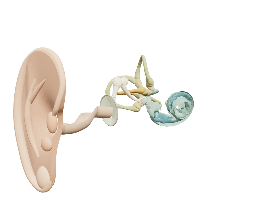
Anatomical right ear: medial inward, superior up, anterior toward the face. Inner-ear fluid spaces retain atlas proportions; outer/middle anatomy is enlarged for study. Choose visible surfaces or use the structure controls. Camera movement changes only the view.
Ear Lab: Complete 2D Reference
Inspect a right-ear teaching model, follow sound transmission, compare physical frequency/amplitude with pitch/loudness and trace an on-screen head turn. The original full hearing and balance article remains below.
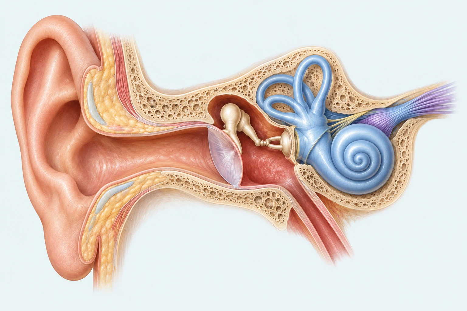
Follow the hearing route from canal to eardrum, bones and cochlea. Inside the cochlea, basilar-membrane motion bends hair-cell structures and initiates neural signaling; the auditory nerve carries those neural messages toward the brain. The visible upper canals belong to balance, not this auditory sequence.
- 1. Ear canal
- The external passage channels sound-pressure changes toward the eardrum.
- 2. Eardrum
- The membrane at the end of the canal vibrates with incoming sound.
- 3. Middle-ear bones
- The connected ossicle chain transfers vibration from the eardrum toward the inner ear.
- 4. Cochlea
- The coiled inner-ear structure supports hair-cell transduction; individual receptors are explained in the text rather than drawn at this scale.
- 5. Auditory nerve
- The neural pathway leaving the cochlear region carries hearing information toward the brain.
- 6. Vestibular canals
- The upper looped canals help detect head rotation for balance; they are not the auditory receptor pathway.
Explore the Ear
- Pinna · Outer Ear
- Function: Collects and channels sound toward the canal.
Location / Role: The visible outer ear sits lateral to the canal. Its folds are a spatial cue; they are not a separate pitch theory.
- Ear Canal · Outer Ear
- Function: Conducts air-pressure changes toward the eardrum.
Location / Role: The canal leads medially inward from the pinna to the eardrum. Air particles move locally around their resting positions.
- Eardrum · Boundary
- Function: Converts pressure changes into mechanical vibration.
Location / Role: The tympanic membrane separates the outer canal from the middle ear. Its attached malleus passes motion into the connected bones.
- Malleus · Middle Ear
- Function: Transfers eardrum motion to the incus.
Location / Role: Its handle attaches to the eardrum; its head joins the incus. The articulated demonstration keeps contacts together.
- Incus · Middle Ear
- Function: Transfers motion between malleus and stapes.
Location / Role: The middle link connects to the malleus and stapes. The joints move as a connected chain, not loose striking bones.
- Stapes & Oval Window · Middle–Inner Boundary
- Function: Transfers mechanical movement into cochlear fluid.
Location / Role: The stapes footplate occupies the oval window at the vestibule. The round window below permits pressure relief; no air enters the cochlea.
- Cochlea · Inner Ear · Hearing
- Function: Houses the mechanical and receptor structures for hearing.
Location / Role: A continuous fluid-filled spiral begins at the base near the oval window and winds to the apex. The opened spiral exposes a schematic basilar ribbon; the cochlear cross-section is simplified.
- Basilar Membrane · Inner Ear · Hearing
- Function: Has frequency-dependent mechanical responses that help encode pitch.
Location / Role: It lies along the cochlear duct; higher frequencies peak near the base and lower frequencies farther toward the apex. The workbench uses a log-frequency teaching map, not measured individual anatomy.
- Hair Cells & Nerve Contact · Inner Ear · Hearing
- Function: Transduce stereocilia deflection into receptor potentials and synaptic output.
Location / Role: Hair cells sit in the organ of Corti above the basilar membrane. The magnified inset shows a shearing structure, stereocilia and separate nerve contacts; spikes occur in nerve fibers.
- Auditory Nerve · Inner Ear → Brain
- Function: Carries action potentials toward auditory brain pathways.
Location / Role: Cochlear fibers join the vestibulocochlear nerve context medially. The route is abbreviated; pressure waves are not traveling down the nerve.
- Horizontal Canal · Inner Ear · Balance
- Function: Detects rotational acceleration mainly in the yaw plane.
Location / Role: It loops from the vestibule in a roughly horizontal plane, with an ampulla near one end. The rightward head-turn example initially increases right-side activity and decreases left-side activity; opposite turning reverses the relationship.
- Anterior Canal · Inner Ear · Balance
- Function: Contributes to detecting rotation in its own plane.
Location / Role: One of the two roughly vertical canals; the three planes are approximately orthogonal. A pure yaw example primarily emphasizes the horizontal canals, not equal activity in all three.
- Posterior Canal · Inner Ear · Balance
- Function: Contributes to detecting rotation in a different plane.
Location / Role: Its vertical plane differs from the anterior canal. Camera rotation changes your view; it does not turn the modeled head.
- Otolith Organs · Inner Ear · Balance
- Function: Contribute gravity, head orientation and linear-acceleration information.
Location / Role: The utricle and saccule lie in the vestibular region between the cochlea and canals. This mode demonstrates canal rotation; it does not attribute all balance information to canals. Kinesthesis also uses receptors outside the ear.
Follow the Sound
- Air Pressure in the Canal. Pressure changes travel through air in the canal. Air particles vibrate locally; they do not travel along the nerve.
- Eardrum Vibrates. Changing pressure moves the eardrum. Its attached malleus begins the middle-ear mechanical chain.
- Ossicles Transfer Motion. The malleus, incus and stapes move through their connected joints. The stapes footplate moves at the oval window.
- Fluid Receives Mechanical Input. Stapes motion at the oval window drives pressure changes in cochlear fluid. The round window permits pressure relief; air does not enter the cochlea.
- Basilar Response Travels. A mechanical response travels from the cochlear base toward the apex. Its largest response depends on frequency: higher near the base, lower farther toward the apex.
- Hair Cells Transduce. Relative motion within the organ of Corti bends stereocilia. Hair-cell receptor potentials change transmitter release to contacting auditory nerve fibers.
- Auditory Fibers Signal. Auditory nerve fibers carry action potentials toward brain pathways. These are neural signals, not pressure waves; hair cells do not grow a new signaling axon.
- Toward Auditory Processing. Brain pathways use the incoming neural activity for auditory processing. This endpoint retains the route cue so you can review the conversion from pressure to mechanical motion to neural information.
Pressure changes in air, mechanical vibration and neural signaling use distinct cues. Motion is enlarged and slowed. Conduction deafness/conductive hearing loss impairs outer/middle-ear transfer; sensorineural deafness/sensorineural hearing loss involves cochlear hair cells or neural pathways. The chosen examples show reduced middle-ear transfer or impaired hair-cell response, not complete silence in every case.
Sound Workbench
One Input, Two Responses
Frequency: 500 Hz · Period: 2.000 ms · Relative pressure amplitude: 0.60.
A temporal pressure trace is a graph, not the shape of air particles traveling into the nerve. In room-temperature air, using 343 m/s, wavelength = speed / frequency = 0.686 m. Frequency relates to pitch; amplitude relates to loudness, which also depends on the listener and frequency. The response curve is an authored teaching approximation.
Changing frequency independently of amplitude moves the modeled peak from basal high-frequency regions toward apical low-frequency regions. Changing amplitude alters pressure/response size without moving the peak. Frequency/wavelength are physical properties related to perceived pitch; amplitude is related to perceived loudness. The physical/perceptual distinction qualifies the CED’s simplified phrasing.
Place theory emphasizes response location; frequency theory emphasizes neural timing for lower-frequency examples; volley theory emphasizes collective timing across fibers. These accounts complement one another. Conservative drawing limits of ≤500 events/s per row and ≤2,000 Hz for volley examples are not official AP cutoffs or a precise physiological model.
Volley Theory
Compare combined timing across illustrative nerve rows: different rows contribute on different cycles. Each shown row has at most 500 events per second in this idealized drawing.
Diagram limits: one row ≤500 events/s; volley examples ≤2,000 Hz. These conservative drawing limits are not official AP cutoffs, individual firing guarantees or an exact allocation among real fibers. Phase locking becomes limited at higher frequencies. The three theories are complementary explanations, not switches in a real ear.
Find the Sound
Source to the left: the nearer ear receives the direct path earlier. Head shadow can also reduce the far-ear level, especially at higher frequencies.
Right minus left arrival: 0.454 ms. Positive means the right ear receives it later.
Authored top-down point-source geometry: ear separation 0.18 m, source radius 2 m, speed 343 m/s in room-temperature air. The illustration is not to scale; distances and timing are calculated model values, not measured listeners. Timing/level cues alone do not uniquely resolve every 3D position.
Balance & Motion
- Begin the Turn. Start from rest, then move the on-screen head through the first part of a yaw turn. The horizontal canal is the relevant example; the page and camera stay still.
- Continue the Turn. As the head continues turning, endolymph initially lags relative to the canal, deflecting the cupula and hair-cell bundles. The response wanes during continued movement.
- Stop the Turn. The head stops at its new angle. Continuing fluid motion briefly produces the opposite relative deflection. Holding an angle does not produce endless rotational activity.
- Settle at the New Angle. The transient response has faded while the modeled head stays at its new angle. Otolith organs contribute gravity and linear-acceleration information separately.
- Turn Complete. The head is held, and the modeled canal response has returned to rest. Vestibular information combines with vision and bodily information; kinesthesis also depends on muscles, tendons and joints outside the ear.
Head Turn → Relative Fluid → Receptor Response
Held angle; transient activity has settled.
One right-ear canal is shown, with the paired left/right relationship explained beside it. This enlarged schematic compresses motion and relaxation time; its deflection is not a physiological measurement. No tilt or linear-acceleration test is simulated.
Semicircular canals contribute rotational information; otolith organs contribute gravity and linear-acceleration information. Vestibular information differs from kinesthesis, which also uses muscles, tendons and joints. No physical turn or balance test is requested.
Sources and Model Assumptions
AP scope: AP Central, current CED Topic 1.6.C and 1.6.G, printed 45–46 / PDF 52–53. Cover: Fall 2025; inner title: Fall 2024. October 2025 clarifications contain no hearing/balance correction.
Mechanisms and anatomy: NIDCD: How Do We Hear?, NIDCD: Balance, UTHealth Auditory Structure/Function, Auditory Processing and Vestibular System. Additional anatomy supports these concepts; it is not a new AP vocabulary requirement.
Original sculpted outer/middle ear joined to selected IE-Map surfaces · Ahmadi et al., 2021, including registered vestibular duct forms from David et al., 2016. CC BY 4.0; adapted by selective conversion, shared coordinate remapping, teaching colors and section openings. The cochlear exterior represents its fluid-space boundary. Inner-ear proportions stay registered; outer/middle anatomy is enlarged for study. This is a combined teaching model, not one whole-ear scan or clinical anatomy. The gold response ribbon follows a source-derived cochlear route; it is not a segmented basilar membrane. The microscopic receptor inset is separately magnified. Shared contacts preserve an enlarged planar ossicle/piston mechanism; real motion is more complex.
Workbench pressure ΔP=A sin(2πft), with milliseconds converted to seconds; T=1/f. The authored peak location is x=1−log₂(f/125)/5 over 125–4,000 Hz, where x=0 is base and x=1 is apex. Response A exp(−½((x−peak)/0.12)²) is a normalized teaching envelope, not a measured tonotopic function or loudness prediction. The displayed peak location is independent of amplitude. Timing rows intentionally omit events above their disclosed display ranges; real phase locking and maximum firing depend on cell, frequency and stimulus. The selected conduction example scales downstream mechanical/receptor cues by 0.28; sensorineural bundle and partial neural cues are reduced separately. These authored visual factors do not estimate a clinical degree of hearing loss. No audio, microphone, hearing test or calibrated output is included.
Localization uses a 2 m source semicircle, ears at ±0.09 m and 343 m/s in room-temperature air; direct-path delay=(distance right−distance left)/343. This excludes diffraction, reverberation and individualized pinna cues. Level differences are explained qualitatively rather than treated as an exact head-shadow measurement. Balance uses a bounded authored turn with transient fluid/cupula cues and a settled held endpoint, not a physiological fluid simulation or dizziness test. Model construction and provenance.
Timing, Place, and Two Ears Provide Different Information
Place theory explains pitch through the location of strongest vibration along the basilar membrane. Different portions respond best to different frequencies; higher frequencies produce strongest responses nearer the base. This account is especially useful for high pitches.
Frequency theory emphasizes firing timed to the sound’s frequency. For lower frequencies, the timing of neural responses can follow individual wave cycles. A single neuron cannot fire at arbitrarily high rates because of its refractory period. Volley theory extends timing-based coding: groups of neurons alternate their firing so the combined pattern can represent frequencies above a single neuron’s maximum rate. Place and timing information can work together; the theories are not three completely separate ears.
Sound localization is identifying where a sound originates. Differences in arrival time and intensity between the ears provide location cues. A sound arriving earlier or more intensely at one ear can help indicate its direction, alongside other cues. Pitch describes what frequency pattern is experienced; localization describes where the source is. A higher-pitched sound is not inherently on the right or left.
| Account | Information used | Key point |
|---|---|---|
Place | Location along basilar membrane | Especially useful for high frequencies. |
Frequency | Timing of firing relative to wave cycles | Useful at lower frequencies. |
Volley | Combined alternating firing of groups | Overcomes some limits of individual firing rates. |
Transmission Problems Differ from Receptor or Nerve Damage
Conduction deafness, or conductive hearing loss, results when sound is not transmitted efficiently through the outer or middle ear. The problem is delivering vibration to the inner-ear receptors. Sensorineural deafness, or sensorineural hearing loss, involves damage to inner-ear hair cells or neural pathways, so receptor transduction or neural signaling is impaired.
Aging and damaging noise exposure can contribute to sensorineural loss. A middle-ear transmission problem and damaged cochlear hair cells may both make hearing difficult, but they disrupt different steps. Use the described mechanism to make the distinction; do not infer the type of loss merely from someone saying sounds seem quieter.
Smell and Taste Detect Chemicals and Jointly Shape Flavor
Olfaction is smell. Airborne molecules interact with receptors in the nose, which initiate neural signals processed through olfactory structures and the brain. Smell is unusual because its initial route to cortex does not first use the thalamic relay that serves most other senses. That does not mean smell never interacts with the thalamus later.
Pheromones are chemical signals released by an organism that can affect other members of the same species through chemical-sensory processing. They are well-established in many animals. This course concept does not establish the existence of a specific human attraction pheromone or validate a commercial fragrance claim.
Gustation is taste. Chemicals dissolved in the mouth stimulate taste-receptor cells in taste buds, which send information through nerves to the brain. Course-listed qualities are sweet (often associated with sugars), sour (acidity), salty (salts), bitter (many potentially aversive compounds), umami (savory qualities associated with glutamate), and oleogustus (a proposed fat-related taste quality). The CED includes oleogustus; its broader scientific classification remains less settled than the five commonly established basic tastes.
People vary in taste sensitivity. Differences in taste-receptor density are related to the distinction among supertasters, medium tasters, and nontasters, with supertasters generally experiencing some tastes more intensely. Nontaster does not mean that the person has no sense of taste. The labels describe comparative sensitivity, and receptor number is not the only possible influence.
Taste and smell interact to produce flavor. With a blocked nose, a person can often still detect basic taste qualities but loses much of the information that differentiates particular foods. The CED’s description of muted taste without smell should be understood in this flavor sense, not as a claim that taste receptors stop responding when olfaction is absent.
Skin Input and Brain Processing Shape Touch and Pain
Skin receptors transduce information about contact, pressure, and temperature into neural signals. Touch experience depends on both this input and processing in brain regions such as the somatosensory cortex. Warm and cold receptor activity contributes to temperature perception. In some combinations, concurrent warm and cold stimulation can create a paradoxical hot or burning experience. That is the useful context for the CED’s warm-plus-cold example; it is not a claim that all heat detection requires equal warm and cold input.
Pain is an experience involving bodily signals and processing in the spinal cord and brain. Gate control theory proposes that transmission of pain-related signals can be modulated, partly at spinal-cord pathways, by other sensory input and descending brain influences. The gate is a model of neural modulation, not a literal door in the skin. Rubbing a bumped area can sometimes reduce experienced pain because non-painful touch input influences those pathways. Attention and expectations can also matter; pain is not simply a meter of tissue damage.
Phantom limb sensation occurs when a person experiences sensation, sometimes pain, in a limb that has been lost. Remaining nerves and central representations can contribute to the experience even when the limb is absent. This shows that bodily experience depends on the nervous system’s processing, not only on receptors in a currently present limb. It does not make the reported pain imaginary.
Pain-related and touch inputs enter spinal pathways; their transmission and the resulting experience depend on spinal and central processing.
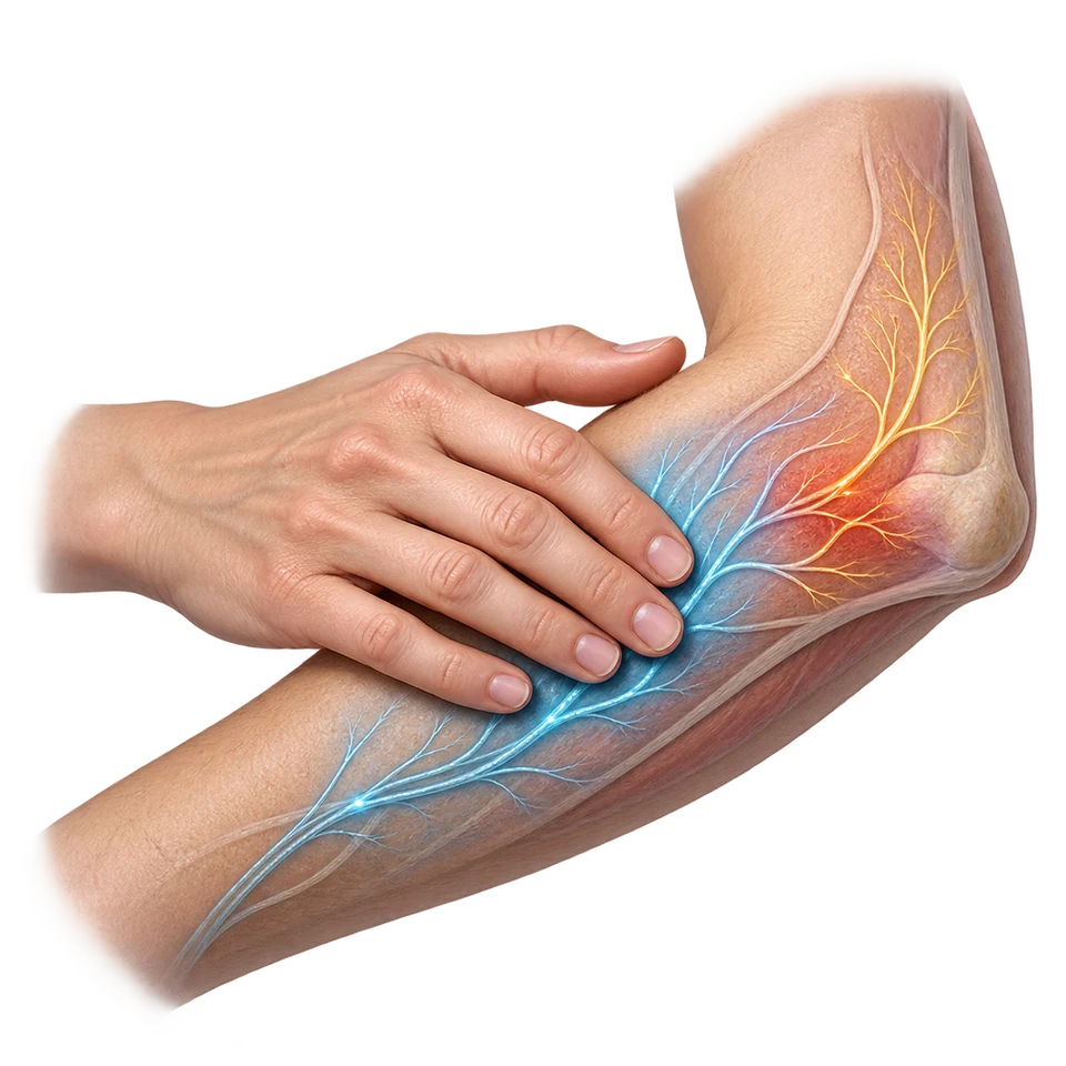 Stage 1
Body Input
Pain-related and non-painful touch signals enter spinal pathways.
- Stage 2
Spinal Modulation
Other input can influence transmission of pain-related signals.
- Stage 3
Brain Processing
Attention, expectations, and descending influences also shape the experience.
Brain → spinal pathways: descending influences can modulate transmission.
A phantom limb can feel painful because neural representations persist.
Knowing Orientation Differs from Knowing Body Movement
The vestibular sense provides information used for balance and spatial orientation. Fluid movement and receptor activity in the semicircular canals of the inner ear signal head rotation; other vestibular structures contribute information about position and linear acceleration. Brain networks integrate these signals with vision and bodily information to maintain balance.
Kinesthesis is the sense of the position and movement of body parts, supported by information from muscles, tendons, and joints. It helps you move in a coordinated way without looking at every limb. Closing your eyes and bringing a finger toward your nose relies on bodily position and movement information. Staying oriented while turning your head strongly engages vestibular information. These senses work together, but balance/orientation and limb movement are distinct contributions.
| Sense | Information | Original illustration |
|---|---|---|
| Vestibular | Head motion and orientation supporting balance | Maintaining balance while turning your head |
| Kinesthetic | Position and movement of body parts | Moving a hand accurately without watching it |
Vestibular information concerns balance and orientation; kinesthetic information concerns body-part position and movement.
Trace the Message. Explain the Consequence.
Follow one connected example: you step off a curb, hear a bicycle bell, turn, and move back. Auditory receptors transduce pressure changes; peripheral sensory pathways relay signals to central networks. The brain interprets input and plans action; somatic motor pathways activate skeletal muscles, while cerebellar systems coordinate movement. Autonomic activity can help mobilize the body. This original scenario combines processes; it does not isolate one region as the sole cause.
If you slept poorly, attention and reaction speed could be affected without changing the bell’s physical frequency. If an auditory pathway were damaged, the difficulty could arise during conduction, transduction, or neural processing. If you learn to respond more efficiently through practice, experience and biological plasticity interact. Trace the altered step before predicting the effect.
Good explanations respect the evidence. A family resemblance does not prove a genetic cause; an fMRI association does not establish exclusive causation; a split-brain response depends on visual field and response channel. A hypothetical dataset illustrates how to read a result, but supplies no published evidence. Carry this distinction between mechanism, application, and inference into every later unit.
Comparisons Worth Being Able to Explain
| Often confused | Distinction to explain |
|---|---|
Nature / nurture | Inherited predispositions / experiences; their interaction shapes outcomes. |
CNS / PNS | Central coordination / pathways linking the CNS with the rest of the body. |
Somatic / autonomic | Skeletal-muscle control / regulation of largely involuntary bodily functions. |
Action potential / synaptic transmission | Electrical change along an axon / chemical signaling across a junction. |
Refractory period / reuptake | Temporary limit on refiring / removal of transmitter from a synapse. |
Agonist / antagonist / reuptake inhibitor | Enhances receptor action / blocks action / reduces reabsorption. |
EEG / fMRI | Recorded electrical patterns / indirect blood-oxygen signals. |
REM behavior disorder / somnambulism | Dream enactment in REM / sleepwalking mainly in deep NREM. |
Absolute threshold / JND | Detecting presence / detecting a change. |
Trichromatic / opponent process | Three cone inputs / comparisons in opponent channels. |
Conduction / sensorineural loss | Vibration transmission / receptor or neural-pathway damage. |
Vestibular / kinesthetic | Balance and orientation / body-part position and movement. |
- Identify the specific scenario detail.
- Explain the relevant mechanism or functional relationship.
- Connect it to the outcome and state the limit of the inference.
Vocabulary + Theory Reference
Organized by official topic. Each term links to its explanation above; chemical messenger and sleep-disorder definitions are repeated here for quick comparison.
1.1 · Heredity & Environment
- Heredity
- Heredity, often called nature, consists of inherited or biologically predisposed characteristics that influence physical traits, behavior, and mental processes.
- Environment
- Environment, often called nurture, includes experiences outside the individual, such as family relationships, education, opportunities, nutrition, and cultural expectations.
- evolutionary perspective
- The evolutionary perspective asks how natural selection could have favored tendencies that supported survival and reproductive success across generations.
- natural selection
- In natural selection, heritable variations associated with greater reproductive success can become more common in a population.
- Eugenics
- Eugenics is the attempt to influence human reproduction according to judgments about supposedly desirable inherited traits. It has been used to justify discrimination and coercion; hereditary differences do not establish differences in human worth.
- Twin studies
- Twin studies compare similarities in identical and fraternal twins, sometimes raised together or apart, to investigate hereditary and environmental contributions to traits. Greater similarity among identical twins can suggest hereditary influence, but differences in shared experiences complicate the inference.
- Family studies
- Family studies examine whether a trait clusters among relatives and whether similarity changes with the degree of biological relationship. Because relatives may share environments as well as heredity, family resemblance alone cannot separate those influences.
- Adoption studies
- Adoption studies compare adopted people with biological and adoptive relatives to investigate heredity and environment. Similarity to biological relatives can suggest hereditary contributions, while similarity to adoptive relatives can suggest environmental contributions; prenatal experience and placement patterns can complicate these conclusions.
1.2 · Nervous System
- central nervous system (CNS)
- The central nervous system (CNS) consists of the brain and spinal cord.
- peripheral nervous system (PNS)
- The peripheral nervous system (PNS) consists of nerves and related neural structures outside the brain and spinal cord. It carries sensory information toward the central nervous system—the brain and spinal cord—and outgoing signals from it to muscles and organs.
- somatic nervous system
- The somatic nervous system is the part of the peripheral nervous system that carries bodily sensory information and controls skeletal muscles. It supports voluntary actions, such as raising a hand, and also participates in involuntary reflexes.
- autonomic nervous system
- The autonomic nervous system is the part of the peripheral nervous system that regulates largely involuntary functions of internal organs and glands, such as heart rate and digestion. Its sympathetic and parasympathetic divisions help adjust bodily activity to changing demands.
- sympathetic division
- The sympathetic division of the autonomic nervous system mobilizes bodily resources for action. During a sudden threat, it can increase heart rate and reduce digestive activity; it also contributes to ongoing bodily regulation.
- parasympathetic division
- The parasympathetic division of the autonomic nervous system supports maintenance, digestion, and recovery. After a challenge, its activity can slow heart rate and support digestion; it continually helps regulate the body rather than simply switching the sympathetic division off.
1.3 · Neurons & Neural Firing
- Neurons
- Neurons are specialized nervous-system cells that receive, process, and transmit information through electrical and chemical signals. For example, a sensory neuron carries information from a skin receptor toward the central nervous system.
- Glial cells
- Glial cells support the nervous system by providing structure, insulation, communication support, and waste transport.
- myelin
- Myelin is insulating material formed by certain glial cells around many axons. It helps electrical signals travel efficiently; damage to myelin can disrupt communication along an axon.
- reflex arc
- A reflex arc is a neural pathway that produces a rapid, automatic response to a stimulus. In a typical withdrawal reflex, sensory neurons carry input to the spinal cord, interneurons connect the signals, and motor neurons activate muscles before conscious awareness of the event.
- sensory neuron
- A sensory neuron carries information from sensory receptors toward the central nervous system. For example, it carries a signal from a heat-sensitive skin receptor toward the spinal cord when you touch a hot surface.
- interneuron
- An interneuron is a neuron within the central nervous system that connects and processes signals among other neurons. In a typical withdrawal reflex, interneurons link incoming sensory information with outgoing motor commands.
- motor neuron
- A motor neuron carries outgoing commands from the central nervous system to an effector, such as a muscle. In a withdrawal reflex, motor neurons activate the muscles that pull a hand away from a hot surface.
- resting potential
- At resting potential, a neuron has an electrical difference across its membrane: the inside is relatively negative.
- Excitatory
- Excitatory describes input that increases a receiving neuron’s likelihood of generating an action potential. It can move the neuron closer to its firing threshold without necessarily causing it to fire by itself.
- inhibitory
- Inhibitory describes input that decreases a receiving neuron’s likelihood of generating an action potential. It can counter excitatory input; inhibitory does not mean harmful or emotionally negative.
- threshold
- A neuron’s threshold is the critical level of membrane depolarization needed to initiate an action potential. Inputs that do not bring the neuron to threshold do not produce that action potential; this neural threshold differs from a sensory detection threshold.
- action potential
- An action potential is a brief electrical impulse that travels along a neuron’s axon after the firing threshold is reached. Each impulse is all-or-none; stronger stimulation can increase firing rate rather than enlarge each impulse.
- depolarization
- Depolarization is a change in membrane voltage that makes the inside of a neuron less negative relative to the outside. During an action potential, sufficiently large depolarization briefly reverses the membrane’s electrical polarity.
- all-or-none principle
- The all-or-none principle means an individual action potential occurs fully once its threshold is reached; it is not a half-sized impulse for a weak stimulus.
- refractory period
- After firing, the refractory period briefly limits the ability to fire again while the membrane returns toward its resting condition.
- synapse
- A synapse is a junction where a neuron communicates with another cell. At a chemical synapse, neurotransmitters released by the sending neuron cross a tiny gap and bind to receptors on the receiving cell.
- reuptake
- In reuptake, a released neurotransmitter is taken back into the sending cell.
- multiple sclerosis
- In multiple sclerosis, damage to myelin in the central nervous system—the brain and spinal cord—disrupts efficient signal transmission, potentially affecting movement, sensation, and other functions.
- myasthenia gravis
- In myasthenia gravis, impaired signaling at the nerve-muscle junction, commonly involving acetylcholine receptors, produces muscle weakness.
- Neurotransmitters
- Neurotransmitters are chemical messengers released by neurons that act on receiving cells at synapses. Their effects depend on the receptors and pathways involved, so one transmitter can contribute to several functions.
- Dopamine
- Dopamine is a neurotransmitter that helps regulate movement, motivation, and reward-related learning. Its effects vary across pathways; reward learning involves more than pleasure alone.
- Serotonin
- Serotonin is a neurotransmitter involved in mood regulation, sleep, and other functions. Mood cannot be reduced to the amount of one transmitter.
- Norepinephrine
- Norepinephrine is a neurotransmitter that supports alertness, attention, and arousal, including responses to challenges.
- Glutamate
- Glutamate is a major excitatory neurotransmitter involved in learning and memory. At many synapses, it increases the receiving neuron’s likelihood of firing.
- GABA
- GABA (gamma-aminobutyric acid) is a major inhibitory neurotransmitter. It reduces the likelihood of firing in receiving neurons and helps regulate neural activity.
- Endorphins
- Endorphins are naturally occurring opioid chemical messengers involved in reducing pain and in related responses. Their pain-reducing role contrasts with substance P’s role in transmitting pain-related signals.
- Substance P
- Substance P is a chemical messenger involved in transmitting pain-related signals. Its role contrasts with that of endorphins, which can reduce pain signaling.
- Acetylcholine
- Acetylcholine is a neurotransmitter that activates skeletal muscles and contributes to attention, learning, and memory. Its effect depends on its receptor and location.
- Hormones
- Hormones are chemical messengers released into the bloodstream that affect cells with suitable receptors.
- Adrenaline
- Adrenaline, also called epinephrine, is a hormone that helps prepare the body for action. It can increase heart rate and mobilize energy during a challenge.
- Leptin
- Leptin is a hormone that signals the availability of stored energy and tends to reduce hunger. Its role is associated with satiety rather than initiating a meal.
- Ghrelin
- Ghrelin is a hormone that helps increase hunger and motivates eating. Its hunger-promoting role contrasts with leptin’s satiety-related signaling.
- Melatonin
- Melatonin is a hormone that helps regulate the timing of the sleep/wake cycle by signaling biological night. It is a timing signal rather than an on-off switch for sleep.
- Oxytocin
- Oxytocin is a hormone involved in childbirth and milk release that also contributes to social bonding. Its social effects depend on context; it is not a universal trust chemical.
- Psychoactive drugs
- Psychoactive drugs change behavior and mental processes by affecting neural communication.
- agonist
- An agonist activates a receptor or enhances the action of a neurotransmitter.
- antagonist
- An antagonist blocks or reduces a neurotransmitter’s action, often by occupying a receptor without activating it in the same way.
- reuptake inhibitor
- A reuptake inhibitor blocks a transporter involved in reabsorbing a transmitter, leaving more of that transmitter available in the synapse for longer.
- Stimulants
- Stimulants, such as caffeine and cocaine, generally increase neural activity and arousal.
- Depressants
- Depressants, such as alcohol, generally reduce neural activity and can impair judgment, coordination, and response speed.
- Hallucinogens
- Hallucinogens are psychoactive drugs that alter perception and cognition, sometimes producing experiences without a corresponding external stimulus. The CED includes marijuana in this category, but its varied effects do not always involve hallucinations.
- Opioids
- Opioids, such as heroin, act on opioid receptors and commonly reduce pain; they can also strongly affect reward and breathing.
- tolerance
- With tolerance, repeated exposure can reduce the effect of the same dose, so a larger dose may be needed to obtain a previous effect.
- Addiction
- Addiction involves compulsive use despite harmful consequences.
- Withdrawal
- Withdrawal refers to adverse symptoms when a substance on which the body has become dependent is reduced or stopped.
1.4 · The Brain
- brain stem
- The brain stem is the region connecting the brain with the spinal cord. It includes the medulla and supports essential functions such as breathing and heart-rate regulation.
- medulla
- The medulla is part of the brain stem that helps regulate vital functions, including breathing and heart rate. Damage there can disrupt these functions even when stored knowledge is not the primary problem.
- reticular activating system
- The reticular activating system helps regulate alertness and the level of wakefulness.
- reward system
- The brain’s reward system is a network that participates in motivation, learning, cognition, and emotion by helping organisms respond to rewarding outcomes.
- cerebellum
- The cerebellum, at the lower rear of the brain, helps coordinate movement and balance and contributes to procedural learning.
- cerebral cortex
- The cerebral cortex is the folded outer layer of the cerebrum, which is divided into left and right hemispheres.
- Corpus callosum
- The corpus callosum is a large bundle of nerve fibers connecting the left and right cerebral hemispheres. It allows information to pass between them; severing it reduces this communication in split-brain research.
- frontal lobes
- At the front, the frontal lobes participate in planning, linguistic processing, and voluntary movement.
- prefrontal cortex
- The prefrontal cortex supports executive functions such as planning, inhibiting an impulsive response, and organizing actions toward a goal.
- motor cortex
- The motor cortex, near the rear of the frontal lobe, helps direct skeletal-muscle movements, largely on the opposite side of the body.
- parietal lobes
- The parietal lobes, near the upper rear portion of the cortex, contribute to integrating and organizing sensory information.
- somatosensory cortex
- The somatosensory cortex receives and processes bodily touch information.
- Association areas
- Association areas are regions of the cerebral cortex that integrate information and support functions such as interpretation, memory, and planning. They are distributed across the cortical lobes rather than confined to the parietal lobes.
- temporal lobes
- The temporal lobes, on the sides of the brain, contribute to hearing and language processing.
- occipital lobes
- The occipital lobes, at the back, are strongly involved in visual processing.
- Thalamus
- The thalamus is a deep brain structure that routes most sensory information toward appropriate cortical regions. Olfaction bypasses this initial relay, although smell can interact with the thalamus later.
- Hypothalamus
- The hypothalamus is a deep brain structure that helps regulate internal balance, including hunger, temperature, and endocrine activity. It communicates with the pituitary gland to help coordinate hormonal responses.
- Pituitary gland
- The pituitary gland is an endocrine gland whose hormone releases operate under hypothalamic influence and help regulate other endocrine functions. It links the brain’s regulation of bodily needs with hormonal activity.
- Hippocampus
- The hippocampus is a brain structure important for forming and consolidating new explicit memories: information about facts and events that can be consciously recalled. It is not the permanent storage site for every type of memory.
- Amygdala
- The amygdala is a brain structure involved in processing emotionally significant information, including threat-related learning. For example, it contributes to learning that a cue predicts danger; it does not produce all emotions by itself.
- split-brain research
- In split-brain research, researchers study people whose corpus callosum has been severed, often as a treatment for severe epilepsy.
- lateralization
- Lateralization is the tendency for some brain functions to be more strongly associated with one hemisphere than the other. Language is often more strongly associated with the left hemisphere, but this does not divide people into logical-left or creative-right personalities.
- Broca’s area
- Broca’s area is a region, typically in the left frontal lobe, that contributes to language production. Damage to this and connected language networks can make speech slow and effortful; production difficulty differs from a general loss of intelligence.
- Wernicke’s area
- Wernicke’s area is a region, typically in the left temporal lobe, that contributes to language comprehension. Damage to this and connected language networks can impair understanding and produce fluent speech that lacks clear meaning.
- aphasia
- Damage to language networks can cause aphasia, an impairment of language.
- left visual field
- The left visual field is the region of visible space to the left of the point of fixation, seen by both eyes. With central fixation, information briefly presented there initially reaches the right cerebral hemisphere; it is not simply input from the left eye.
- right visual field
- The right visual field is the region of visible space to the right of the point of fixation, seen by both eyes. With central fixation, information briefly presented there initially reaches the left cerebral hemisphere; it is not simply input from the right eye.
- Brain plasticity
- Brain plasticity is the capacity to change connections and patterns of functioning through development, experience, and sometimes recovery after injury.
- EEG (electroencephalogram)
- An EEG (electroencephalogram) records electrical activity using scalp electrodes.
- fMRI (functional magnetic resonance imaging)
- fMRI (functional magnetic resonance imaging) uses changes related to blood oxygenation as an indirect indicator of activity and helps locate regions associated with a task.
- Case studies
- Case studies examine an individual in depth and can reveal valuable patterns after unusual injuries; they are limited in generalization.
- Lesioning
- Lesioning damages or removes tissue and can help establish how disrupted structures affect functioning, but planned harmful lesions in healthy humans are not an ethical experimental method.
1.5 · Sleep
- Consciousness
- Consciousness is awareness of thoughts, feelings, behavior, and internal or external events.
- circadian rhythm
- A circadian rhythm is an approximately 24-hour biological cycle.
- Jet lag
- Jet lag follows a rapid shift across time zones when internal timing has not yet aligned with local day and night.
- Shift work
- Shift work is work scheduled outside a conventional daytime schedule, including overnight or rotating shifts. It can require wakefulness during biological night and sleep during biological day, disrupting alignment with the circadian rhythm.
- NREM (non-rapid eye movement) sleep
- Non-rapid eye movement (NREM) sleep comprises stages N1, N2, and N3, with brain-activity patterns that differ from rapid eye movement (REM) sleep. It ranges from light transitional sleep to deep slow-wave sleep; dreaming can occur during both NREM and REM sleep.
- N1
- N1 is the transition into light sleep, with slower activity than alert wakefulness; hypnagogic sensations, such as a falling sensation, may occur as a person enters it.
- hypnagogic sensations
- Hypnagogic sensations are vivid sensory experiences that can occur while falling asleep, especially during the transition into N1 sleep. Feeling as though you are falling while drifting off is one example.
- N2
- N2 is a stage of non-rapid eye movement (NREM) sleep characterized by sleep spindles, brief bursts of rhythmic electrical activity, and K-complexes, large brief changes in recorded brain electrical activity. These patterns distinguish N2 from the transition into N1 and the deep slow-wave activity of N3.
- N3
- N3 is the deepest stage of non-rapid eye movement (NREM) sleep, characterized by slow, high-amplitude waves in recordings of brain electrical activity. It is especially common early in the night and differs from rapid eye movement (REM) sleep’s more wake-like brain activity.
- REM (rapid eye movement) sleep
- REM (rapid eye movement) sleep involves relatively active, low-amplitude mixed-frequency brain activity resembling wakefulness, while most voluntary skeletal muscles are strongly relaxed.
- paradoxical sleep
- Paradoxical sleep is another name for rapid eye movement (REM) sleep. The paradox is that recorded brain electrical activity resembles wakefulness while most voluntary skeletal muscles are strongly inhibited; eye movements and essential bodily functions continue.
- REM rebound
- REM rebound is an increase in rapid eye movement (REM) sleep that can follow REM deprivation. Subsequent sleep contains more REM sleep relative to the person’s previous pattern, rather than simply more total sleep.
- Activation-synthesis theory
- Activation-synthesis theory proposes that the brain constructs a meaningful dream from internally generated activity during sleep.
- Consolidation theory
- Consolidation theory of dreaming proposes that dreams are related to the processing and strengthening of information from waking experience during sleep. A dream incorporating a recent experience is compatible with the theory, but does not by itself prove that consolidation occurred.
- Memory consolidation
- Memory consolidation is the process by which new memories become more stable and organized.
- Restoration theory
- Restoration theory proposes that sleep helps replenish resources and supports repair and recovery after waking activity.
- Insomnia
- Persistent difficulty falling asleep, staying asleep, or obtaining satisfactory sleep, with resulting daytime effects. It differs from choosing not to allow enough time for sleep.
- Narcolepsy
- Narcolepsy is a disorder involving marked daytime sleepiness and recurrent episodes of sleep; features associated with rapid eye movement (REM) sleep can intrude into waking life. It is not ordinary tiredness after a late night.
- REM sleep behavior disorder
- REM sleep behavior disorder involves disruption of the normal muscle inhibition during rapid eye movement (REM) sleep, allowing a person to physically act out dream-related movements.
- Sleep apnea
- Repeated interruptions or reductions in breathing during sleep that disrupt sleep and can cause daytime sleepiness.
- Somnambulism
- Somnambulism is sleepwalking, usually associated with deep non-rapid eye movement (NREM) sleep. It differs from dream enactment during rapid eye movement (REM) sleep when normal muscle inhibition is disrupted.
1.6 · Sensation
- Sensation
- Sensation is detecting stimulation and converting it into signals the nervous system can use.
- Transduction
- Transduction is the conversion from physical or chemical stimulation into neural signals.
- Perception
- Perception is organizing and interpreting sensory information.
- absolute threshold
- The absolute threshold is the minimum intensity detected on at least 50% of trials under specified conditions.
- just-noticeable difference (JND)
- The just-noticeable difference (JND), or difference threshold, is the smallest detectable difference between two stimuli under specified testing conditions. Detecting a small increase in a weight illustrates a difference threshold; detecting a weight at all concerns an absolute threshold.
- Weber’s law
- Weber’s law proposes that the change needed for detection is approximately a constant proportion of the starting intensity, within a relevant range.
- Sensory adaptation
- Sensory adaptation is reduced sensitivity during sustained, unchanging stimulation, such as noticing a steady odor less after entering a room.
- Sensory interaction
- Sensory interaction occurs when information from one sense influences an experience produced with another. For example, a food’s aroma changes its flavor even when taste receptors still detect sweetness.
- Synesthesia
- Synesthesia is a consistent experience in which stimulation in one sensory or cognitive system is accompanied by an experience in another, such as sounds eliciting color experiences.
- retina
- The retina is the light-sensitive layer at the back of the eye. Its rods and cones convert light into signals that retinal circuits process and send toward the brain through the optic nerve.
- blind spot
- The blind spot is the retinal location where the optic nerve exits; it lacks photoreceptors.
- lens
- The lens is a transparent structure in the eye that helps focus light on the retina. Changes in its shape adjust focus for objects at different distances.
- accommodation
- Accommodation in vision is the process by which the eye’s lens changes shape to focus light from objects at different distances onto the retina. This optical adjustment differs from changing a mental schema, also called accommodation in development.
- Nearsightedness
- Nearsightedness means distant objects are hard to focus sharply, commonly because light focuses in front of the retina.
- Farsightedness
- Farsightedness involves difficulty with near focus, commonly because light would focus behind the retina without sufficient compensation.
- Rods
- Rods are especially useful in dim light and support sensitivity to light, shapes, and movement without color discrimination.
- Cones
- Cones support color vision and fine detail in sufficient light.
- fovea
- The fovea is a small central region of the retina with a high concentration of cones, supporting sharp central vision and fine detail. Directly focusing on small print brings its image onto the fovea.
- Trichromatic theory
- Trichromatic theory explains color vision through the combined responses of three cone types sensitive to overlapping ranges of short, medium, and long wavelengths. Their relative activity encodes color at the receptor level; later pathways also use opponent processing.
- Opponent-process theory
- Opponent-process theory explains further processing in opponent channels: red versus green, blue versus yellow, and black versus white.
- afterimage
- An afterimage is a visual experience that persists after the original stimulus is removed. A negative color afterimage can appear in an opponent color, such as greenish after looking at red, because visual pathways adapt to sustained stimulation.
- Color vision deficiency
- Color vision deficiency involves altered color processing, often because a cone class functions differently or is absent, and sometimes because of other visual-pathway problems.
- Dichromatism
- Dichromatism is a form of color vision deficiency with two functioning cone classes rather than the usual three. This limits some color distinctions, such as red-green distinctions in common forms, without necessarily eliminating all color vision.
- monochromatism
- Monochromatism is a form of color vision deficiency in which only one cone class functions, or ordinary cone-based color discrimination is absent. With insufficient differences between cone signals to encode hues, color vision is severely limited or absent.
- Prosopagnosia
- Prosopagnosia, or face blindness, involves difficulty recognizing faces even when other visual abilities are relatively intact.
- blindsight
- In blindsight, a person with damage affecting primary visual processing may respond to some visual information in a region reported as unseen, without normal conscious visual awareness there.
- Frequency
- Frequency is the number of sound-wave cycles per second, measured in hertz (Hz). Higher frequency generally corresponds to higher perceived pitch; frequency is a physical property of the wave rather than the subjective experience.
- pitch
- Pitch is the perceived highness or lowness of a sound, closely related to its frequency. A high-frequency whistle generally has a higher pitch than a low-frequency rumble; pitch differs from loudness.
- Amplitude
- Amplitude is the size of a sound wave’s pressure variation. Greater amplitude generally produces greater perceived loudness, while leaving frequency and pitch potentially unchanged.
- loudness
- Loudness is the perceived intensity of a sound. It is related to wave amplitude but also depends on frequency and the listener; turning up a recording can make it louder without raising its pitch.
- eardrum
- The eardrum is a thin membrane that vibrates in response to sound waves entering the ear canal. Its movement passes vibrations to the middle-ear bones, helping transmit sound toward the inner ear.
- cochlea
- The cochlea is the fluid-filled, coiled structure of the inner ear that contains hearing receptors. Incoming vibrations move its basilar membrane and stimulate hair cells, converting sound-related motion into neural signals.
- basilar membrane
- The basilar membrane is a structure inside the cochlea that moves in response to sound. Different locations respond most strongly to different frequencies, and its motion bends hair-cell structures that initiate auditory transduction.
- Place theory
- Place theory explains pitch through the location of strongest vibration along the basilar membrane.
- Frequency theory
- Frequency theory explains pitch through the timing of neural firing that follows a sound wave’s frequency. It works best for low frequencies, because a single neuron cannot fire fast enough to match arbitrarily high sound frequencies.
- Volley theory
- Volley theory extends timing-based coding: groups of neurons alternate their firing so the combined pattern can represent frequencies above a single neuron’s maximum rate.
- Sound localization
- Sound localization is identifying where a sound originates. Differences in arrival time and intensity between the ears provide cues to direction; localization describes where a sound comes from rather than its pitch.
- Conduction deafness
- Conduction deafness, or conductive hearing loss, results when sound is not transmitted efficiently through the outer or middle ear.
- Sensorineural deafness
- Sensorineural deafness, or sensorineural hearing loss, involves damage to inner-ear hair cells or neural pathways, so receptor transduction or neural signaling is impaired.
- Olfaction
- Olfaction is the sense of smell, produced when odor molecules stimulate receptors in the nose and their signals are processed by the brain. Unlike most senses, its initial route to cortex does not first pass through the thalamus.
- Pheromones
- Pheromones are chemical signals released by an organism that influence other members of the same species. They are well established in many animals, but that does not establish a specific human attraction pheromone or validate fragrance advertising.
- Gustation
- Gustation is the sense of taste, produced when chemicals dissolved in the mouth stimulate taste-receptor cells. It detects basic taste qualities, whereas flavor also incorporates smell and other sensory information.
- sweet
- Sweet is the taste quality commonly elicited by sugars and some other compounds, including certain sweeteners. The sweetness of sugar dissolved in water illustrates this quality without depending on a food’s aroma.
- sour
- Sour is the taste quality associated with acidity. The sharp taste of lemon juice illustrates sourness; it differs from bitter taste rather than serving as another name for it.
- salty
- Salty is the taste quality commonly elicited by dissolved salts, especially sodium chloride. The taste of lightly salted water illustrates saltiness rather than a smell-based flavor.
- bitter
- Bitter is a taste quality elicited by many compounds, including caffeine. Unsweetened coffee can illustrate bitterness, although its full flavor also involves aroma; a bitter taste does not by itself establish that a substance is poisonous.
- umami
- Umami is a savory taste quality associated especially with glutamate. It contributes to the savory taste of broth and differs from simply tasting salt.
- oleogustus
- Oleogustus is the proposed taste quality of certain free fatty acids, distinct from the creamy or greasy texture of fatty foods. It is included in the AP Psychology CED, although its status as a basic taste is less settled than that of the five commonly established taste qualities.
- supertasters
- Supertasters are people who experience certain tastes, especially some bitter compounds, unusually intensely compared with other people. Taste-receptor density can contribute, but sensitivity is not determined by receptor number alone and does not imply superior ability to identify every flavor.
- medium tasters
- Medium tasters are people with intermediate sensitivity to certain taste stimuli, commonly assessed with bitter-taste tests. They respond more strongly than nontasters and less strongly than supertasters to those test compounds; this is a comparison of sensitivity rather than a separate sense.
- nontasters
- Nontasters are people with low sensitivity to certain test compounds, commonly bitter substances used to classify taste responsiveness. They can still taste other substances; nontaster does not mean a complete absence of the sense of taste.
- flavor
- Flavor is the combined experience of a food or drink produced by taste, smell, and other sensations such as texture and temperature. A blocked nose can reduce a food’s distinctive flavor even while sweetness or saltiness remains detectable.
- Pain
- Pain is an unpleasant sensory and emotional experience associated with actual or potential tissue damage, or resembling that experience. It depends on nervous-system processing and is not a direct meter of injury; phantom limb pain can occur even when the limb is absent.
- Gate control theory
- Gate control theory proposes that transmission of pain-related signals can be modulated, partly at spinal-cord pathways, by other sensory input and descending brain influences.
- Phantom limb sensation
- Phantom limb sensation occurs when a person experiences sensation, sometimes pain, in a limb that has been lost.
- vestibular sense
- The vestibular sense provides information used for balance and spatial orientation.
- semicircular canals
- Fluid movement and receptor activity in the semicircular canals of the inner ear signal head rotation; other vestibular structures contribute information about position and linear acceleration.
- Kinesthesis
- Kinesthesis is the sense of the position and movement of body parts, supported by information from muscles, tendons, and joints.
Sources + Scope
Original Get Psyched teaching prose, examples, and diagrams grounded in the currently linked College Board AP Psychology CED, Fall 2025 cover edition, printed pp. 29–46 (PDF indexes 35–52), and the October 2025 clarifications. Retrieved October 8, 2026. Printed pages and zero-based PDF indexes are different.
The October corrections reflected here are “all-or-none principle,” reticular activating system alertness, and “hypnagogic.” The inner title page still says Fall 2024; this review uses the file currently linked by AP Central with the Fall 2025 cover and corrections.
Scientific diagrams show simplified relationships, not experimental evidence. All worked scenarios and the plotted detection and sleep examples are invented for teaching. No published finding is attributed to an invented participant.
Accuracy Notes
The CED groups cortex and deeper brain structures together; this guide distinguishes their locations while teaching all listed structures. It distinguishes sound frequency from perceived pitch, basic taste from combined flavor, and receptor agonism from overall excitation. Supporting factual checks include NIH’s hearing explanation and NIH’s sleep-stage explanation.
Stay Inside the Required Scope
Detailed genetics and the sodium-potassium pump are excluded. Study only the eight listed neurotransmitters, five hormones, and five sleep disorders as the Unit 1 required lists. Endocrine-gland detail is excluded except for the pituitary connection. Psychoanalytic dream theory is excluded here. Descriptive diagram labels support functional understanding; this unit emphasizes applying functions rather than a separate structure-identification drill.